11.2.1 POJA-L3194+3191+3175+3176+3205+3307+3174
Title: Ganglion cells and nerve fibers
Description:
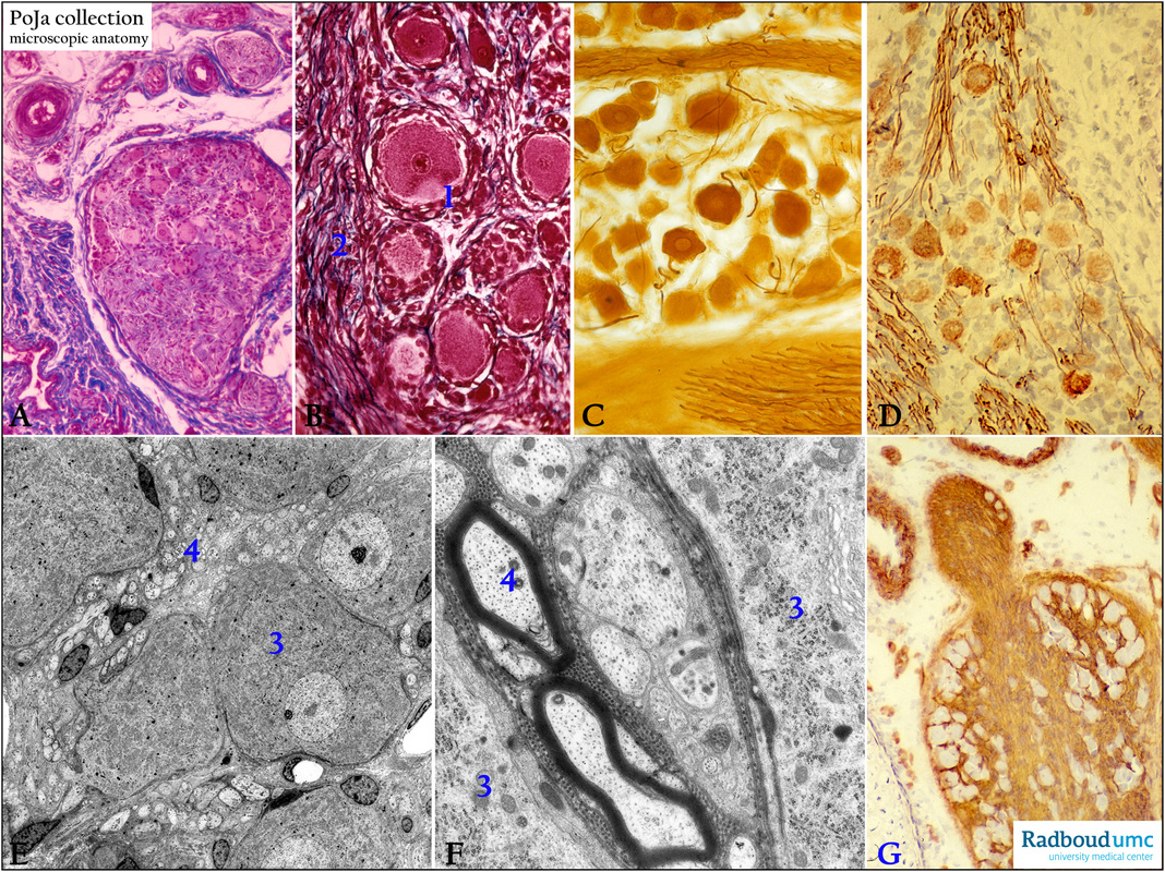
(A): Stain Mallory trichrome, human. Peripheral sympathetic neuron originating from the prostatic plexus and lower part of the hypogastric plexus. Note the presence of large ganglion cells surrounded by satellite or glial cells, as well as the myelinated axons.
(B): Stain Azan, human. Spinal ganglion cells with surrounding amphicytes. Note the clear axon hillock (1) and the myelinated axons (2).
(C): Silver stain, neonate human. Large spinal ganglion cells and thin thread-like axons.
(D): Immunoperoxidase staining with AEC and antibodies against neurofilaments (NF) showing the positive cochlear ganglion cells and
the axons, 1d postnatal rat.
(E): Electron micrograph showing a survey of ganglion cells in the spinal ganglion, rat. (3) Perikaryon ganglion cell with numerous ribosomes. (4) Unmyelinated axons and small nuclei of Schwann cells.
(F): Electron micrograph of the periphery of two spinal ganglion cells, rabbit. (3) Nissl body and Golgi area, details of two myelinated axons (4) close to unmyelinated ones. Note the cross-sectioned neurotubules in the axons.
(G): Immunoperoxidase staining with AEC and antibodies against laminin, spinal ganglion, 1d postnatal rat. The laminin-positive part of
the tissue embeds the cross-sectioned small white, unstained ganglion cells.
Keywords/Mesh: nervous tissue, ganglion cell, Schwann cell, axon, myelin, Nissl body, neurofilament, laminin, histology, electron microscopy, POJA collection

