12.2.3 POJA-La0100+L3385+3384+La0102
Title: Auditory tube in the middle ear
Description:
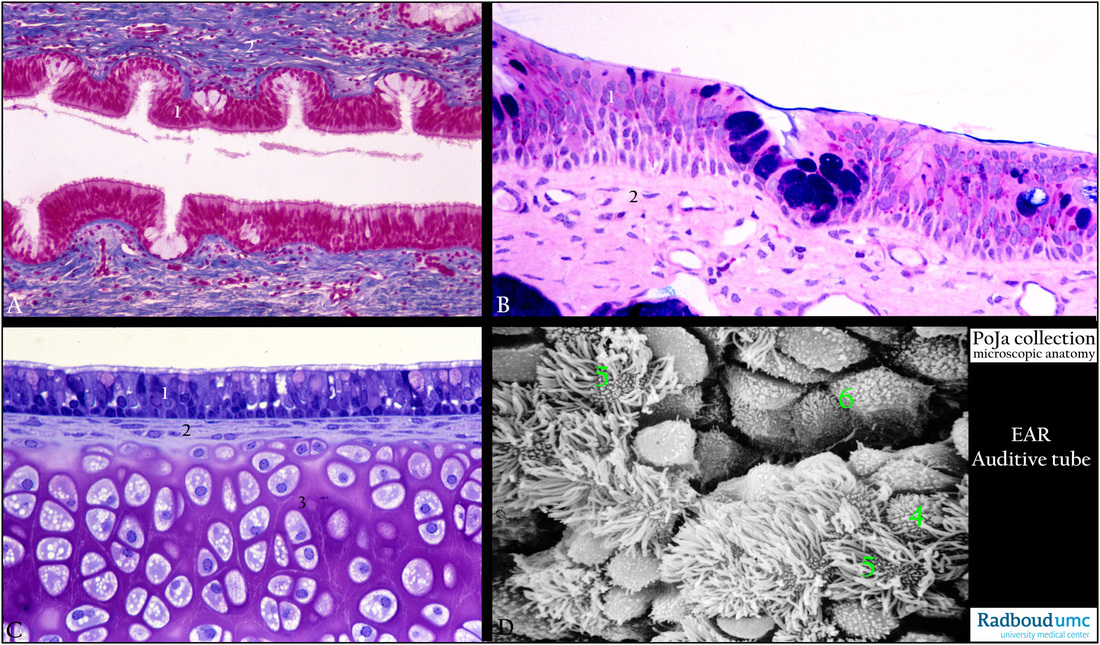
(A): Lining epithelium of auditory tube (pharyngotympanic duct or eustachian tube), stain Azan, human.
(B): Lining epithelium of auditory tube, stain alcian blue-PAS, semi-thin plastic section, guinea pig.
(C): Lining epithelium and cartilage of auditory tube, stain toluidine blue, cartilage is stained pink, semi-thin plastic section, guinea pig.
The pictures in (A, B, C) show that the wall of the tube is covered with a lining epithelium and stroma and reinforced with hyaline cartilage.
(1) The lining consists of a multilayered ciliated epithelium (respiratory type).
Goblet cells are present both as individual cells (white, (A), deep blue- or pink- stained (B, C) in the lining as well as intraepithelial glands.
(2) The proper lamina with close to the cartilage a thin layer of perichondrial cells that give rise to the chondroblasts.
(3) The cartilage shows so-called isogenous cell groups of chondroblasts surrounded by a territorial matrix.
After division the daughter cells remain within the same lacuna. Between the territorial matrices deep pink interterritorial matrix (in C)
is characteristic in the hyaline cartilage (see 15.1 Locomotor system: Cartilage: 15.1 POJA -L7005+7004 Hyaline cartilage)
(D): Lining epithelium of auditory tube after tubal obstruction, scanning electron micrograph, rat.
Normally the eustachian tube (pharyngotympanic duct) is closed but opens at short intervals (e.g. swallowing) equalising pressure
in the middle ear cavity with the external air pressure.
Tubal obstructions are generally considered as important factors for the pathogenesis of chronic otitis media with the formation of effusion.
Development of cell hypertrophy and increased secretory activity is observed when an eustachian tubal obstruction occurs
upon induction of sterile middle ear effusions. As a result the surface of the ciliated epithelium shows initial (D, 4) and advanced (D, 5) stages of neogenesis of ciliated cells. Goblet cells are also activated (D, 6) and are studded with short plump microvilli.
Keywords/Mesh: middle ear, eustachian tube, auditory tube, respiratory epithelium, ciliated epithelium, effusion, histology, pathology, POJA collection

