15.1 POJA-L7014+7015 Hyaline cartilage with asbestos transformation (polarisation)
15.1 POJA-L7014+7015 Hyaline cartilage with asbestos transformation (polarisation)
Title: Hyaline cartilage with asbestos transformation (polarisation)
Description: Haematoxylin-eosin staining of human hyaline cartilage.
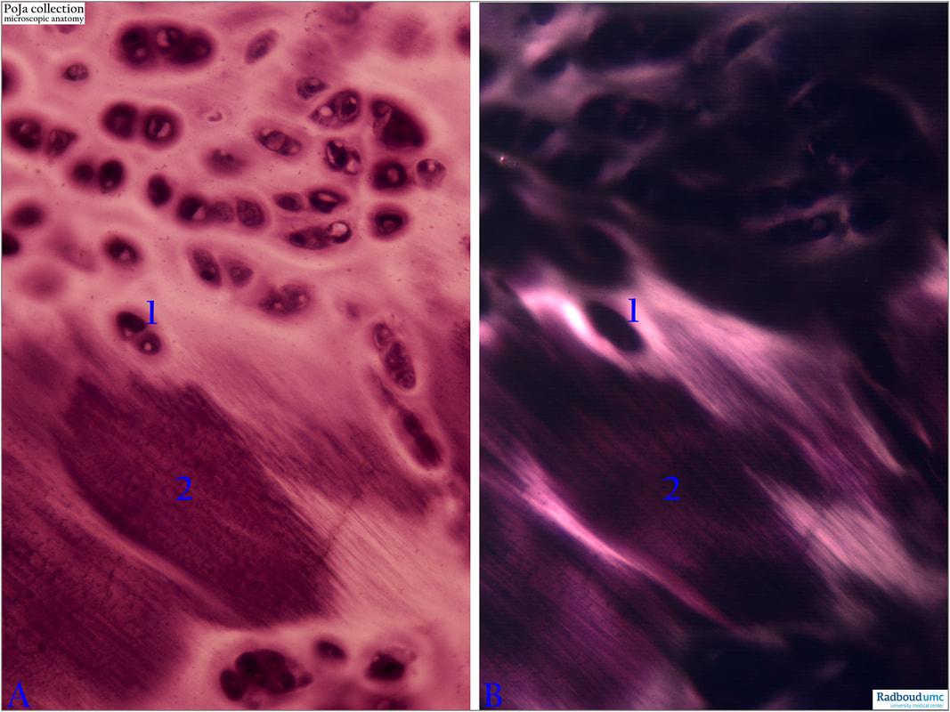
(A): Normal light microscopy. (B): Polarisation microscopy of the same section.
(1): Isogenous group of two chondrocytes embedded in territorial matrix. Upper half of the slide contains numerous isogenous islets of chondrocytes.
(2): The collagen fibrils in the interterritorial area are becoming visible upon gradual demasking during senescence. The fibrils in (2) are stained dark or light depending on the polarisation angle and whether they run all in one direction or cross each other in different angles.
Background:
The numbers (1) and (2) (in both A and B) point to exactly the same isogenic chondrocytes (1) and the area (2) of asbestos transformation (or amianthoid changes). Light microscopically amianthoid changes (or transformation) formerly called asbestoid degeneration (or transformation) refers to a focal conversion of the hyaline matrix of cartilage into a characteristic fibrous structure that consists of straight parallelly arranged argyrophilic fibres without any chondrocytes islets. The ‘unmasking’ of collagenous fibrils results in a greater visibility of collagenous components. Amianthoid changes are observed during the second decade of life in man and is remarkable in that ageing is accompanied by increased order.
The increasing anisotropy of the collagen in amianthoid transformation has been confirmed by Hukins et al. in 1976 using high angle X-ray diffraction that provided evidence that amianthoid change, occurring during ageing of costal cartilage, corresponds to a transformation from an isotropic to a marked anisotropic distribution of collagen fibrils.
Low-angle x-ray diffraction and electron microscopy show that the fibrils have the customary 60 -70 nanometre axial periodicity. Normal diameters of collagen fibrils are 20-200 nm. Electron microscopy shows that wide amianthoid collagen fibrils consist of smaller parallel fibrils fused together (see Ghadially 2013).
See also:
References
Keywords/Mesh: locomotor system, cartilage, hyaline, polarisation microscopy, matrix, isogenous chondrocyte, collagen fibre, asbestos degeneration, amianthoid transformation, histology, POJA collection
Title: Hyaline cartilage with asbestos transformation (polarisation)
Description: Haematoxylin-eosin staining of human hyaline cartilage.
(A): Normal light microscopy. (B): Polarisation microscopy of the same section.
(1): Isogenous group of two chondrocytes embedded in territorial matrix. Upper half of the slide contains numerous isogenous islets of chondrocytes.
(2): The collagen fibrils in the interterritorial area are becoming visible upon gradual demasking during senescence. The fibrils in (2) are stained dark or light depending on the polarisation angle and whether they run all in one direction or cross each other in different angles.
Background:
The numbers (1) and (2) (in both A and B) point to exactly the same isogenic chondrocytes (1) and the area (2) of asbestos transformation (or amianthoid changes). Light microscopically amianthoid changes (or transformation) formerly called asbestoid degeneration (or transformation) refers to a focal conversion of the hyaline matrix of cartilage into a characteristic fibrous structure that consists of straight parallelly arranged argyrophilic fibres without any chondrocytes islets. The ‘unmasking’ of collagenous fibrils results in a greater visibility of collagenous components. Amianthoid changes are observed during the second decade of life in man and is remarkable in that ageing is accompanied by increased order.
The increasing anisotropy of the collagen in amianthoid transformation has been confirmed by Hukins et al. in 1976 using high angle X-ray diffraction that provided evidence that amianthoid change, occurring during ageing of costal cartilage, corresponds to a transformation from an isotropic to a marked anisotropic distribution of collagen fibrils.
Low-angle x-ray diffraction and electron microscopy show that the fibrils have the customary 60 -70 nanometre axial periodicity. Normal diameters of collagen fibrils are 20-200 nm. Electron microscopy shows that wide amianthoid collagen fibrils consist of smaller parallel fibrils fused together (see Ghadially 2013).
See also:
- 15.1 POJA-L7002+7012+7013 Hyaline cartilage visualised with polarisation microscopy
- 15.1 POJA-L7006+7007+7011 Hyaline cartilage
References
- Hukins, D.W.L., Knight, D.P., Woodhead-Galloway, J. Amianthoid change: orientation of normal collagen fibrils during aging Science 194, 622-624, 1976 DOI: 10.1126/science.982030
- Ultrastructural pathology of the cell and matrix F.N. Ghadially 4th edition 2013, Butterworth-Heinemann ISBN-13: 978-0750698566
Keywords/Mesh: locomotor system, cartilage, hyaline, polarisation microscopy, matrix, isogenous chondrocyte, collagen fibre, asbestos degeneration, amianthoid transformation, histology, POJA collection

