
9.7 POJA-L2911+2913+3459.
Glomus caroticum (carotid body) (II) in human
9.7 POJA-L2911+2913+3459
Title: Glomus caroticum (carotid body) (II) in human
Description:
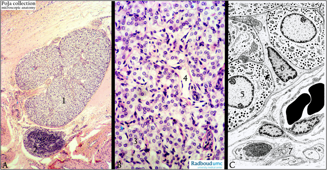
(A, B): Carotid body, stain hematoxylin-eosin. (A, 1) Glomus caroticum adjacent to a lymph node (A, 2).
(B, 3) Shows the main cells or the glomus type I cells. (B, 4) Capillary.
(C): Electron microscopy scheme. (5) Main cell. (6) Supporting cell or glomus type II cell.
(7) Axon endings and a myelophage process (dark).
Background: The glomus or paraganglion is a peripheral chemoreceptor. The main cells produce biogenic amines, they are derived from the Anlage of the sympathetic system (i.e. neural crest)
Keywords/Mesh: glomus caroticum, carotid body, paraganglion, neuroendocrine cell, histology, electron microscopy, POJA collection
Title: Glomus caroticum (carotid body) (II) in human
Description:
(A, B): Carotid body, stain hematoxylin-eosin. (A, 1) Glomus caroticum adjacent to a lymph node (A, 2).
(B, 3) Shows the main cells or the glomus type I cells. (B, 4) Capillary.
(C): Electron microscopy scheme. (5) Main cell. (6) Supporting cell or glomus type II cell.
(7) Axon endings and a myelophage process (dark).
Background: The glomus or paraganglion is a peripheral chemoreceptor. The main cells produce biogenic amines, they are derived from the Anlage of the sympathetic system (i.e. neural crest)
Keywords/Mesh: glomus caroticum, carotid body, paraganglion, neuroendocrine cell, histology, electron microscopy, POJA collection