14.1 POJA-L6132+6050+6051+6052 Cross sections of skeletal muscle
14.1 POJA-L6132+6050+6051+6052 Cross sections of skeletal muscle
Title: Cross sections of skeletal muscle
Description:
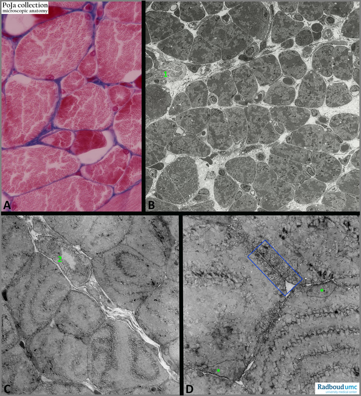
(A): Cross section with Cohnheim areas, Azan staining, human.
The polygonal shaped myofibres are well defined by blue stained endomysium. Within the large myofibres areas of irregularly demarcated fields of myofibrils are seen. These areas are the so-called areas of Cohnheim. It is assumed to be an artefact due to shrinkage. Apart from the large myofibres several smaller ones and a deep-red hypercontracted myofibre are present.
(B): Electron microscopy, a low survey of cross sectioned myofibres mouse. At (1) part of an arteriole and its branch. Between the myofibres the endomysium shows capillaries and free cells. At this low magnification the light areas in the myofibres represent the bundles of myofilaments, the denser areas stand for cross sectioned regions of the Z-lines.
(C, D): Higher magnification, mouse. In (C) an arteriole at (2), light grey areas are the sectioned myofilaments (mainly from the A-band). The curved trails made up of rhomboid structures are sectioned Z-line areas, and the variable curving depends on the state of contraction of individual myofibres.
In (D) two peripheral nuclei (*) and also many light swollen mitochondria (rectangle). Most larger mitochondria are located at the periphery and due to individual contraction circumstances sections of the Z-line areas are depicted as solitary dense spots or as half-curved trails.
Keywords/Mesh: locomotor system, skeletal muscle, striated muscle, Cohnheim area, A-band, I-band, Z-line, mitochondrion, electron microscopy, histology, POJA-collection

