14.5 POJA-L6078+6079+6125 and 14.5 POJA-L6350 Myotendinous junctions in skeletal muscle
|
14.5 POJA-L6078+6079+6125 and 14.5 POJA-L6350 Myotendinous junctions in skeletal muscle
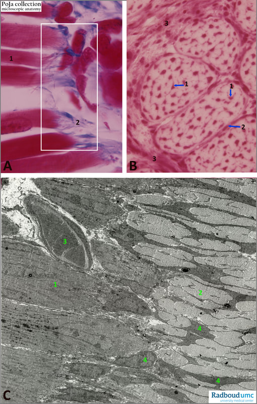
Title: Myotendinous junctions in skeletal muscle Description: The scheme (14.5 POJA-L6350) shows the macro area of the myotendinous junction. 14.5 POJA-L6078+6079+6125 (A): Longitudinal section, Azan stain, human. (1) Muscle fibres of which the surrounding endomysial connective tissue continues in the (blue) tendon (2). Rectangle is the transition zone comparable to (C). (B): Cross section through the tendon (white, pure collagen), Nuclear red stain, human foetus. (1): Tenocytes, or tendon cells with the morphology of fibroblast but with wing-like processes. They build up the ECM and collagen fibres. The tenoblasts or stem cells are usually located in the capsule or endotenon (2) around a primary bundle of tenocytes, as well as in the epitenon or in the paratenon (3), being the capsule around several primary bundles, at the outside of the tendon. |
(C): Idem, electron micrograph of the transition area, mouse. (1): Muscle fibres with banding pattern (2): Tendon fibres. (4): Tenocytes. (3): Capillary with erythrocytes.
Background:
The tendon attaches skeletal muscle to bone and in similar way ligaments attach bone to bone. Tendon consists of fascicles that are well organised and regularly arranged, on the other hand fascicles of ligaments are less properly organised.
A tendon is composed of bundles or fascicles of dense regular connective tissue. The primary fibre bundle is composed of numerous parallelly arranged collagen fibres and are enveloped by the endotenon, within the bundle the so-called tenocytes (or tendinocytes) are located squeezed between the collagen fibres. These cells are specialised internal fibroblasts with wing-like processes and in light microscopy their nuclei are visible in a linear row in longitudinal sections of the tendon. The epitenon surrounds a group of fascicles followed by an outer skirt of connective tissue with vascularisation (paratenon). Epitenon and paratenon are also referred as peritenon.
See also
Keywords/Mesh: locomotor system, skeletal muscle, tendon, ligament, dense fibrous connective tissue, dense regular connective tissue, collagen fibril, tenocyte, tendinocyte, endotenon, epitenon, paratenon, peritenon, electron microscopy, histology, POJA collection
Background:
The tendon attaches skeletal muscle to bone and in similar way ligaments attach bone to bone. Tendon consists of fascicles that are well organised and regularly arranged, on the other hand fascicles of ligaments are less properly organised.
A tendon is composed of bundles or fascicles of dense regular connective tissue. The primary fibre bundle is composed of numerous parallelly arranged collagen fibres and are enveloped by the endotenon, within the bundle the so-called tenocytes (or tendinocytes) are located squeezed between the collagen fibres. These cells are specialised internal fibroblasts with wing-like processes and in light microscopy their nuclei are visible in a linear row in longitudinal sections of the tendon. The epitenon surrounds a group of fascicles followed by an outer skirt of connective tissue with vascularisation (paratenon). Epitenon and paratenon are also referred as peritenon.
See also
- 14.5 POJA-L6121+6081+6123+6082 Myotendinous junctions in skeletal muscle
- 14.5. POJA-L6124+6080+6122 Skeletal Muscle-Tendon linkage
- 14.5 POJA-L6351 Anatomy of transition of muscles into tendon
Keywords/Mesh: locomotor system, skeletal muscle, tendon, ligament, dense fibrous connective tissue, dense regular connective tissue, collagen fibril, tenocyte, tendinocyte, endotenon, epitenon, paratenon, peritenon, electron microscopy, histology, POJA collection


