
14.6 POJA-L6321+6320+6322 Autophagy in myopathy (human)
14.6 POJA-L6321+6320+6322 Autophagy in myopathy
|
14.6 POJA-L6321+6320+6322 Autophagy in myopathy (human)
(By courtesy of L. Eshuis BSc Section Electron microscopy, Department of Pathology, Radboud university medical center, Nijmegen, the Netherlands)
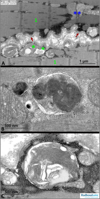
Title: Autophagy in myopathy (human) Description: (A): In normal and myopathic muscle fibres autophagic vacuoles are always present in varying quantities. Two myofibres (1) are separated by interstitium with collagen fibrils. The lamina basalis (red arrows) is apposed to the sarcolemma. The peripherally located mitochondria are distorted and degenerated into membranous whorls (*). Note at (**) large and swollen mitochondria with extended cristae, a myelin whorl is already present. Dense stained granules are glycogen particles. (B): Various types of autophagic vacuoles formed by nonspecific processes. The smallest dense vacuole is surrounded by a halo, the second largest dense one containing some glycogen granules. The large autophagic vacuole is a distinct example of nonspecific macroautophagy demarcated by a single membrane with internalised remnants of cristae and membranes of mitochondria (C): In a dense stained glycogen-rich area a distinct membrane-limited autophagic vacuole with whorling membrane remnants. Background: Autophagy is a specialised cellular pathway in which organelles, cytoplasmic proteins are degraded in the lysosomal compartment. Autophagic vacuoles are the result of a nonspecific process in which an organelle or a cytoplasmic portion is enclosed by a double or multilamellar membrane of the endoplasmic reticulum (so-called isolation membrane) forming the vacuole or autophagosome. Lysosomal enzymes are delivered into the autophagosome and subsequently it matures into a lysosome. Keywords/Mesh: locomotor system, skeletal muscle, striated muscle, autophagic vacuole, macroautophagy, lysosome, mitochondrion, glycogen, membrane whorl, electron microscopy, POJA collection |