
4.2.1 POJA-L3699+3702+3718+3709. Histochemical characterization of liver cells (human, rat)
4.2.1 POJA-L3699+3702+3718+3709
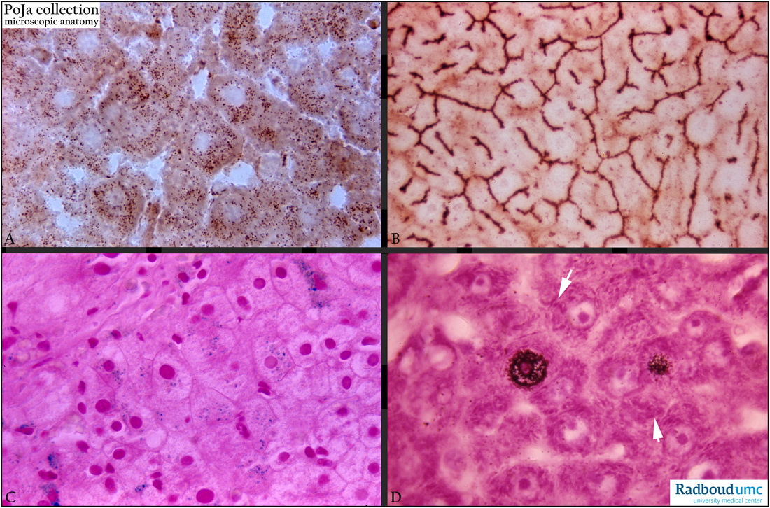
Title: Histochemical characterization of liver cells (human, rat)
Description:
(A) Catalase-DAB stained in phase contrast (rat). Endogenous catalase activity is shown in brown peroxisomes.
(B) Adenosine triphosphatase (Gomori) (rat). The brown-stained ATPase activity marks the localization of bile canaliculi between the liver cells.
(C) Perl's iron stain (human). The blue stained grains represent stored iron in the liver cells.
(D) Autoradiography of tritium thymidine labeling in DNA-replicating nuclei (dark dots) of liver cells, counterstained with pyronin to localize RNA in polyribosomes in RER aggregates (arrows) (mouse).
Keywords/Mesh: liver, histochemistry, thymidine labeling, ATPase, bile canaliculi, Perl’s stain (iron), peroxidase, histology, POJA collection