13.1 POJA-L4650
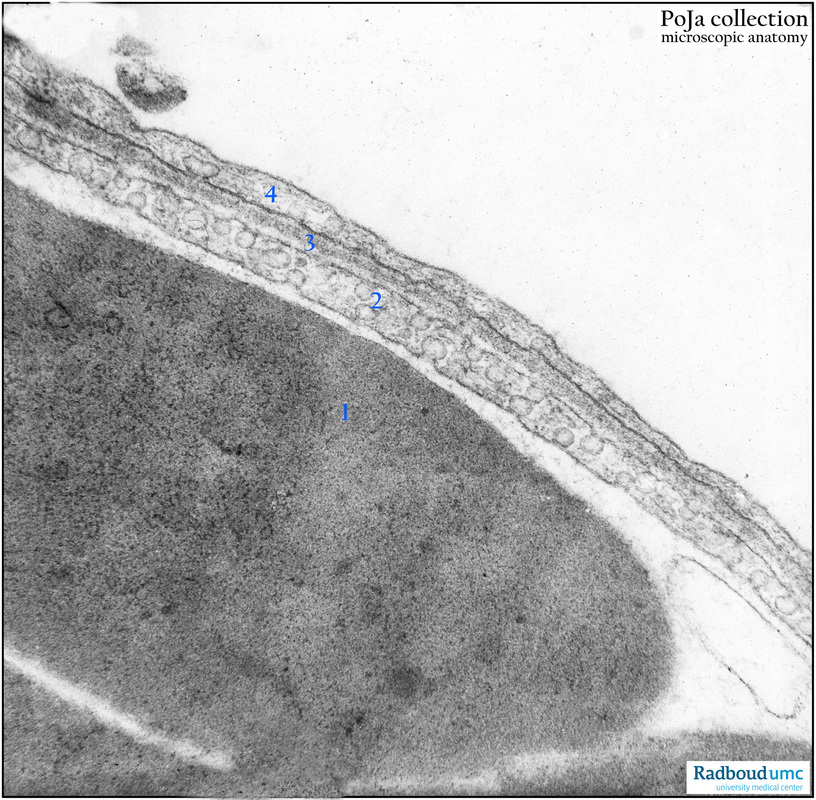
Title: Alveolar capillary in the lung (dog)
Description:
Electron micrograph of a lung capillary. (1) Erythrocyte in the capillary lumen. (2) Slender extension of the endothelial cell with numerous pinocytotic vesicles. (3) Common or fused lamina basalis between capillary and type I alveolar cell (type I pneumocyte) constituting the Air-Blood Barrier for the gaseous exchange. (4) Type I pneumocyte.
Background: Capillaries with continuous endothelium and an uninterrupted basal membrane are most common, i.e. non-fenestrated somatic capillaries. Lung capillaries adjacent to the alveolar spaces share fused basal laminae of the endothelium and the alveolar epithelium (Air-Blood-Barrier).
Keywords/Mesh: cardiovascular system, vascularisation, lung, alveolus, aveolar cell, pneumocyte, capillary, endothelium, pinocytosis, Air-Blood barrier, histology, electron microscopy, POJA collection

