5.4.2 POJA-L2436+La0083+2368+2490+2352+5018+5019+5020
Title: Macula densa in kidney IX
Description:
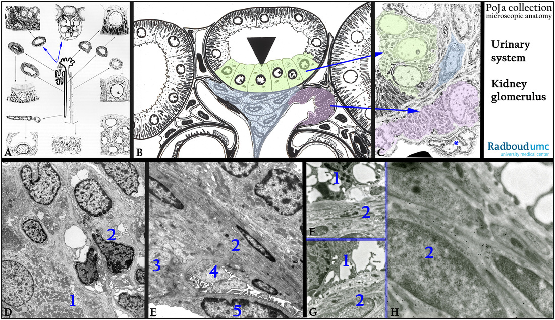
(A): Nephron, scheme, human. The blue arrows at (6) point to the glomerulus and the macula densa as the final part of cortical thick ascending limb (before the distal convolution) that is closely apposed to the glomerulus.
(B): Macula densa (light green), electron microscopy scheme, human. The JG cells with renin granules are painted pink.
The extramesangial lacis cells are bluish.
(C): Detailed scheme of (B) showing that the JG cells are modified smooth muscle cells with renin granules and located just below
an endothelial cell (*) of a capillary. One of the three lacis cells is painted in blue.
(D, E): Macula densa cells (1) and lacis cells (2), electron microscopy, rat (D), rabbit (E).
In (E) the extraglomerular mesangium cells or lacis cells (2) are continuous with the intraglomerular mesangium cells (3).
(4) Podocyte and (5) endothelial cell.
(F-H): Electron micrograph of immunogold labeling of heparan sulfate with gold particles linked antibodies (4C3) against HS, rat.
HS is shown to be localized in the basement membrane matrix between the lacis cells (2) and the macula densa cells (1).
Please zoom in with your viewer to see the detailed structures!!
Keywords/Mesh: urinary system, kidney, glomerulus, juxtaglomerular apparatus, macula densa, JG cell, lacis cell, heparan sulfate, histology, electron microscopy, POJA collection

