11.2 POJA-L3243+3225+3240+3241+3245+3242
Title: Peripheral nerves
Description:
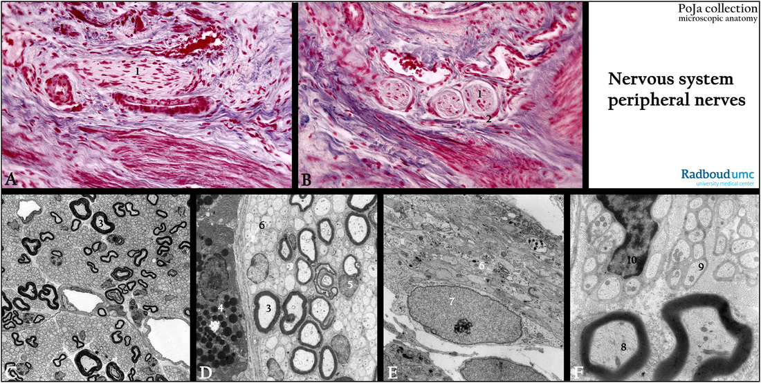
(A): Stain Azan, human. The nerve fiber bundle (1) is embedded in connective tissue (blue) in the vicinity of smooth muscle fibers.
The spindle shaped nuclei are part of the Schwann cells or fibroblasts in the endoneurium.
(B): Mallory stain, human. Cross-sectioned nerve bundles (1) with each a perineurium sheath (2) in cavernous body of penis.
(C): Electron micrograph, golden hamster. Survey of electron-dense myelinated nerve fibers (3) located in peripheral lung tissue.
(D): Electron micrograph, golden hamster. Electron-dense myelinated nerve fibers (3) near exocrine pancreas tissue (4). Note cell of Schwann (5) for myelinated axon, as well as unmyelinated ones (6).
(E): Electron micrograph, human fetus. Subcutaneous tissue in the limb shows (6) unmyelinated axons (7) nucleus of Schwann cell.
(F): Electron micrograph, golden hamster. A cross-section through the area of a principal bronchus. (8) Myelinated nerve fiber. (9) Unmyelinated nerve fibers. (10) Nucleus of the Schwann cell enclosing several nerve fibers.
Keywords/Mesh: nervous tissue, peripheral nerve fiber, Schwann cell, histology, electron microscopy, POJA collection

