4.2.1 POJA-L-3655+3656
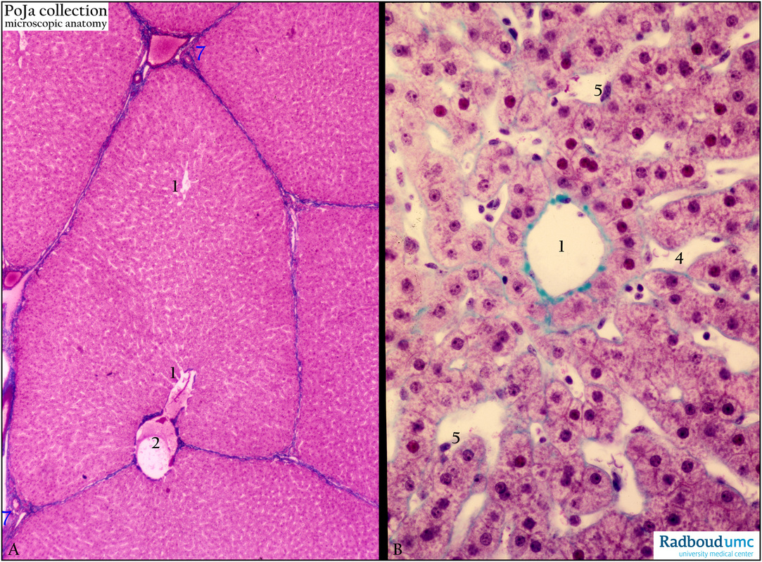
Title: Liver with central veins (pig, human)
Description: Stain: (A) Azan; (B) Trichrome-Goldner.
(A) Survey (pig) and (B) detail (human) with central veins (1). Note that in (B) the “central” vein has a thin rim of connective tissue (4). The central vein in (A) passes to the sublobular vein (2). (5) Kupffer cell in the sinusoid. (7) Periportal area or portal triad (Kiernan). Note that the liver of the pig (A) is clearly divided in units (hepatic lobules) or hepatons by thin (blue) connective tissue septa, while the human liver has a much less defined hepaton structure (not shown in B).
Keywords/Mesh: liver, hepaton, central vein, periportal area, histology, POJA collection

