POJA-L-2927+2934
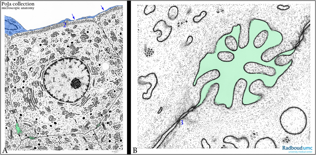
Title: Scheme liver cell and intercellular bile canaliculus (human)
Description: (A): Electron micrograph scheme of a liver cell (1) with microvilli (2) extended in the space of Disse. The covering sinusoid cell (endothelial cell) is stained blue and its long slender extensions are fenestrated (arrows↘). The liver cell is well equipped with RER, Golgi profiles and mitochondria as well as glycogen. Adjacent cells have formed bile canaliculi (green) shown enlarged in (B). The canaliculus is studded with microvilli and is sealed off with tight junctions (3).
Keywords/Mesh: liver, bile canaliculus, space of Disse, sinusoid cell, scheme, electron microscopy, histology, POJA collection

