
12.2.4.1 POJA-L3431+2625+
3433+3425+3400+2626
Stria vascularis I in the cochlea of the inner ear
12.2.4.1 POJA-L3431+2625+3433+3425+3400+2626
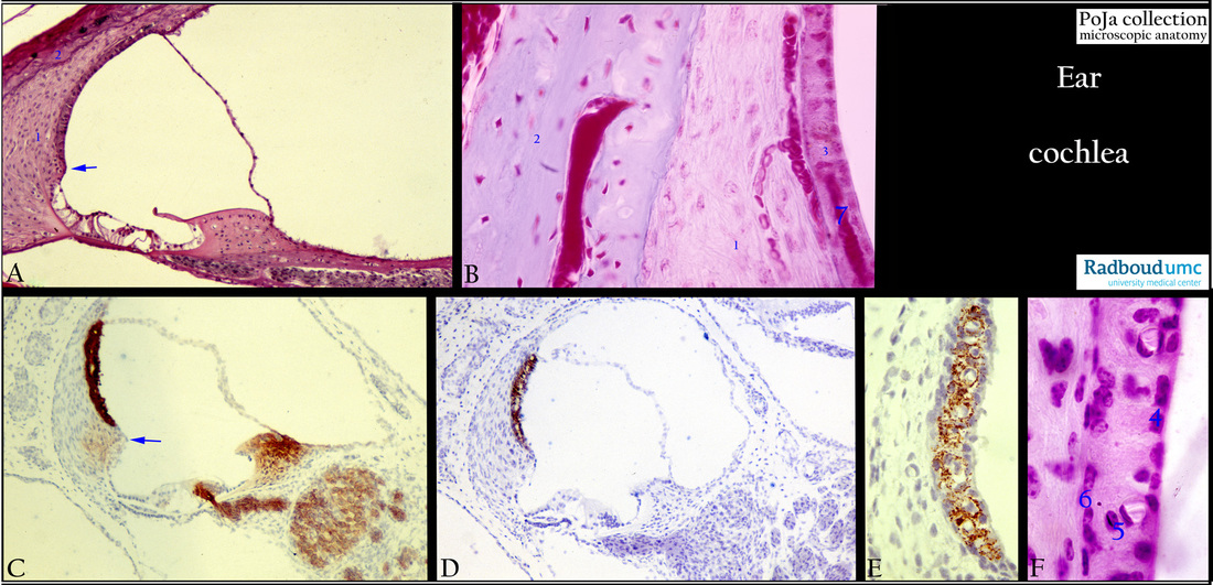
Title: Stria vascularis I in the cochlea of the inner ear
Description:
(A): Ductus cochlearis, stain Haematoxylin-azophloxine, guinea pig.
Adjacent to the stria vascularis is the spiral prominence (A, C arrow) as a thickened region
of the outer spiral ligament. It is the transition of the stria vascularis into the cells of the organ of Corti.
(B): Stria vascularis, stain Azan, guinea pig.
The outer spiral ligament (A, 1; B, 1) is the modified thickened endosteum of the outer wall of the bony cochlear duct (A, 2; B, 2).
It is composed of a moderately dense connective tissue with a lot of blood vessels and numerous close-connected fibroblasts (B).
The stria vascularis is that part of the spiral ligament lined by vascularised specialised stratified epithelium (B, 3).
(C): Ductus cochlearis, immunoperoxidase staining with AEC and antibody against Na+/K+-ATPase, 10 days postnatal rat.
The stria vascularis produces endolymph identical to the intracellular fluid and its ionic composition, and
is involved in the K+ homeostasis. Hence the high Na+/K+-ATPase activity and potassium channels of the intermediate cells (C, see F).
The latter play a crucial role in the production and maintenance of +90mV endocochlear potential and ionic environment
necessary for the marginal cells (see F4) in the cochear duct.
(D): Ductus cochlearis, immunoperoxidase staining with AEC and the antibody NKI beteb, 10 days postnatal, rat. See detail in (E)
(E): Stria vascularis, immunoperoxidase staining with AEC and the antibody NKI beteb, intermediate cells,10 days postnatal, rat.
The antibody NKI-beteb is an anti-melanoma associated antigen and recognise cells of the melanocytic lineage i.e.
the intermediate cells stain distinctly positive (in D, E).
(F): Stria vascularis, stain Haematoxylin-eosin, guinea pig.
The epithelium (F) consists of marginal cells (4) facing the lumen of the cochlear duct, melanocytes (5) as intermediate cells and
a layer of basal cells (6) as well as 2 capillaries (with open lumen). Intraepithelial capillaries (see B, 7) are characteristic for the stria vascularis.
Keywords/Mesh: inner ear, cochlea, stria vascularis, intermediate cell, endolymph, K+ homeostasis, NKI-beteb, melanin, histology,
POJA collection