10.5 POJA-L2170+2174+2177+2178
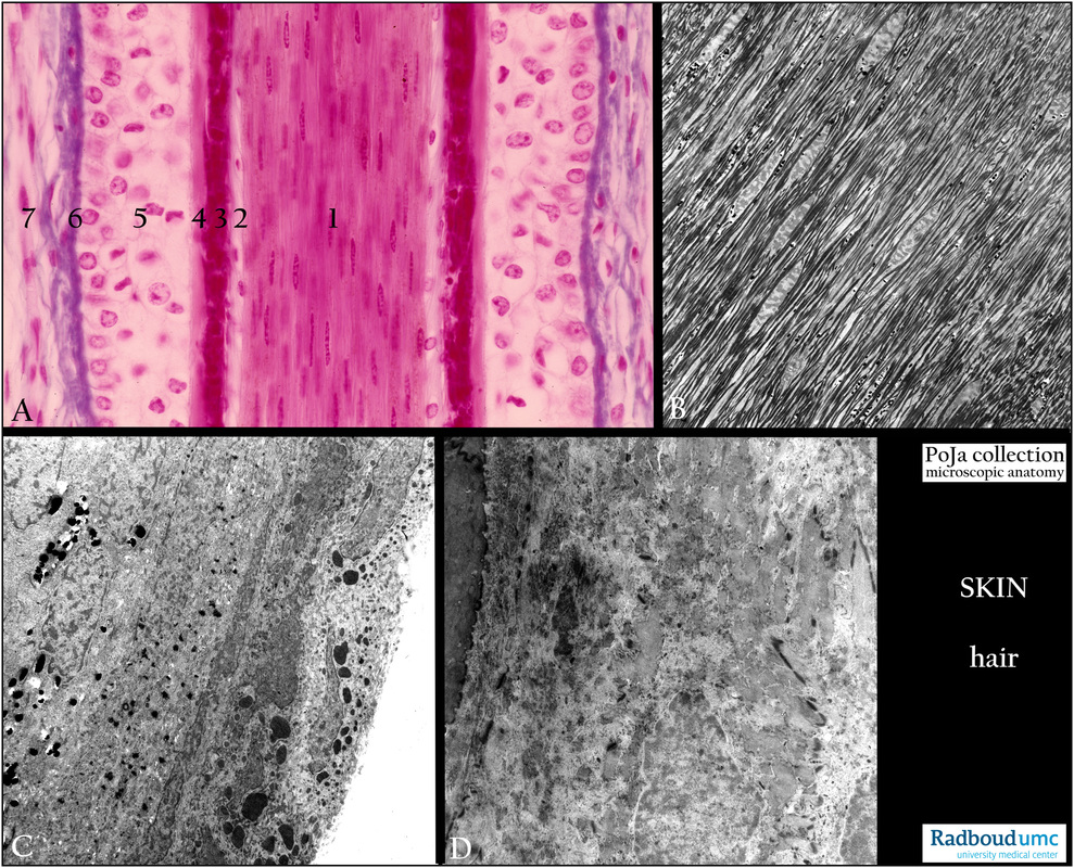
Title: Different sections V of the human hair
Description:
(A): Longitudinal section hair (head), stain Azan.
(1) Hair medulla. (5) External root sheath.
(2) Cuticle of the sheath. (6) Glass membrane.
(3) Huxley’s layer, part of internal root sheath. (7) Connective tissue sheath.
(4) Henle’s layer, part of internal root sheath.
(B - D): Electron micrographs and details of hair shaft.
(B): The central epithelial cells of the cortex are parallel localized and show electron-light nuclei with compact bundles of electron-dense cytokeratin filaments (tonofilaments) in their cytoplasms.
(C): Most left the peripheral part of the cortex with epithelial cells filled with electron-dense pigment granules and strands of tonofilament bundles (cytokeratin). Subsequently a thin cell layer (filled with an elongated electron-greyish structure) of the cuticle, closely apposed to the cell layers of Huxley with the large electron-dense keratohyaline granules (part of the internal root sheath).
(D): From the left to the right the electron-dense area of the corneal layer followed by the granular layer(par of the external root sheath). (The cells here appear a bit degenerated).
Keywords/Mesh: skin, hair, histology, electron microscopy, POJA collection

