5.4.3 POJA-L2502+2432+La0065+2503+2505+2506+2507
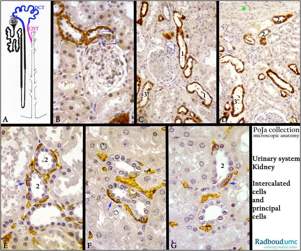
Title: Distal convoluted tubules (DCT) and connecting tubules (CNT) (VIII) in the kidney
Description:
(A): Scheme nephron highlighting in blue the DCT, and in pink the CNT, human.
(B): Kidney cortex, immunoperoxidase staining with DAB and anti-parvalbumin antibodies, rat. The DCT’s are positive while the
macula densa (blue rectangle) remains negative.
(C, D): Kidney cortices, immunoperoxidase staining with DAB and anti-Ca-binding protein-28 kDa antibodies, human.
The macula densa (blue rectangle) remains negative while the (1) distal convoluted tubule (DCT) is positive. (D, 2) subcapsular (*) connecting tubule (CNT) is positive. In (C, D 3) the cells of the cortical collecting duct (CCD) are positive except for the intercalated
cells (IC).
(E - G): Kidney cortices, immunoperoxidase staining with DAB and antibodies against erythrocyte anion exchanger 1 (AE1, AE2, AE3)
or Band 3, human. This transport protein mediates the exchange of chloride for bicarbonate. The protein is present in the basolateral surface of the α-intercalated cell in the collecting duct of the kidney. The α-intercalated cell is the principal acid secreting cell of the
kidney, involved in the acidification of the urine. The blue arrows point to the position of the positive cells.
Note that in (E, G 2) a connecting tubule (CNT) shows several positive streaks of basal labyrinth in the cells.
The erythrocytes in the capillaries are stained stained positively due to endogenous peroxidase.
Keywords/Mesh: urinary system, kidney, cortex, macula densa, DCT, CNT, CCD, intercalated cell, Ca-binding protein, Band 3 protein, histology, POJA collection

