4.2.1 POJA-L3670+3671+3672
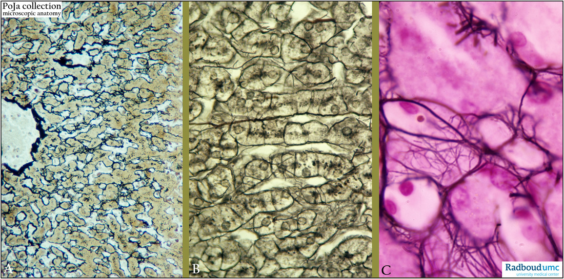
Title: The reticular network around liver parenchymal cells (human)
Description: Stain: (A) Movat. (B) Gomori. (C) Laguesse.
The liver parenchymal cells are surrounded with a “wire network” of argyrophilic fibers, called reticular fibers that are running in the space of Disse, together with collagen I, III and V meshwork fibers. In fact this meshwork stabilizes the sinusoid canals between the liver cells. They are visualized by three different staining procedures (A, B and C).
Keywords/Mesh: liver, space of Disse, reticular fibers, sinusoid, histology, POJA collection

