
7.1 POJA-L1263+1261+1334. Ultrastructure oocyte-follicular cells in ovary (gerbil)
7.1 POJA-L1263+1261+1334
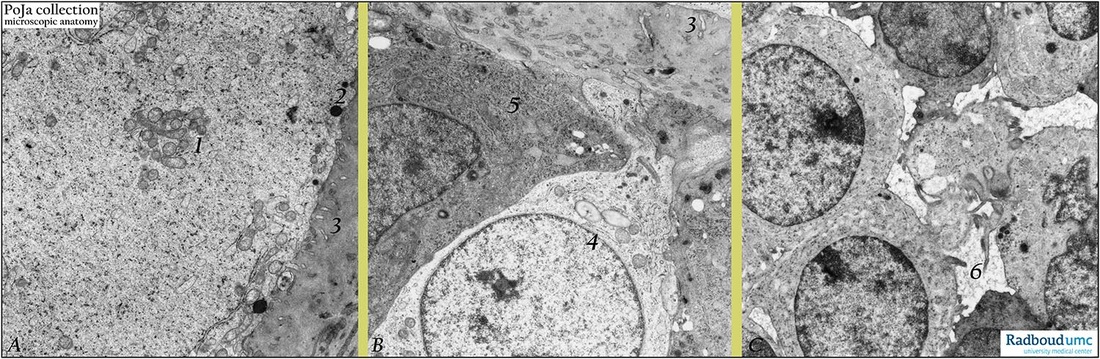
Title: Ultrastructure oocyte-follicular cells in ovary (gerbil)
Description: Electron microscopy (gerbil).
(A): Secondary follicle (early type 4) with part of oocyte. The periphery of the ooplasm contains aggregated mitochondria (1) mixed with small vesicles, few profiles of rough endoplasmic reticulum. Free ribosomes and denser-stained glycogen particles are also present. Close to oolemma few electron-dense cortical granules (2). Short microvilli of oocyte are present within the electron-grey amorphous basal lamina-like zona pellucida (3).
(B): Electron-light (4) and -darker (5) granulosa cells of corona radiata with interzonal microvilli present in the electron-grey basal lamina-like zona pellucida. Oocyte microvilli at (3).
(C): Early type 4 secondary follicle with junctioned electron-light and –grey granulosa cells. Ultra structurally distinct intercellular clefts (6) filled with liquor are already present between granulosa cells. However, they are hardly detectable at light-microscopical level.
Background: It is already assumed that communication between oocyte and granulosa cells is bidirectional. The interplay is essential for development of oocyte as well as for granulosa cells. This so-called oocyte-granulosa regulatory loop is very important for differentiation of normal follicles and for a competent oocyte to undergo fertilization. Although gonadotrophins are necessary the differentiation of granulosa cell phenotypes the oocyte is probably the dominant factor to determine direction of differentiation and associated functions of granulosa cells. Differences in electron-density between neighboring granulosa cells reflect the amounts of intracytoplasmic organelles indicating varying functional activities on time. The zona pellucida plays an important role in the fertilization process; it is composed of a.o. glycoproteins ZPGp1, ZPGp2, ZPGp3 and ZPGp4. It is known that capacitation (occurring as sperm remains one up to six hours in the female genital tract) includes the process of loss of spermatozoal glycocalyx as well as vesiculation of the acrosome of spermatozoon. In the binding process of spermatozoa oligosaccharides linked to ZPGp3 interact with sperm receptors. It appears that only acrosome-reacted spermatozoon can interact with ZPGp3. The cortical reaction refers to the exocytosis of cortical granules (produced by Golgi areas) discharging their contents (mixtures of proteases) in the zona pellucida shortly after sperm-oocyte fusion. Proteases alter the pellucidal structure resulting in a specific transformation of the zona pellucida that penetration by other capacitated spermatozoa (polyspermy) is prevented (so-called zona reaction). The cortical granules from the Golgi areas migrate to the periphery of the oocyte fuse with the oocyte plasma membrane (oolemma). The cortical reaction refers to the exocytosis of these granules discharging their contents (mixtures of proteases) in the ZP shortly after sperm-oocyte fusion. Proteases alter the pellucidal structure resulting in a specific transformation of the ZP that penetration by other capacitated spermatozoa is prevented (so-called zona reaction).
Keywords/Mesh: female genitalia, ovary, ovarian follicle, oocyte , zona pellucida, granulosa cells, histology, POJA collection