
13.1 POJA-L4660+La-0310
Mid-sized vein (human)
13.1 POJA-L4660+La-0310
Title: Mid-sized vein (human)
Description:
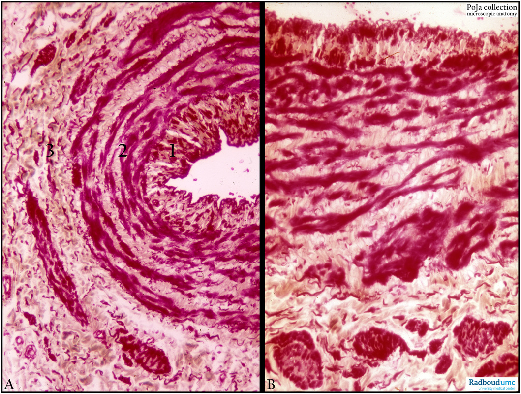
(A-B): Survey and detail of a medium-sized vein (branch of femoral vein), resorcin-fuchsin and light green. These smaller veins show in different areas of the wall longitudinal arranged smooth muscle cells (SMC) in intima and adventitia. The latter generally shows a variable thickness over the whole length of the vessel. The media appears usually well-developed. At (1) the intima with longitudinal arranged SMCs, media (2) with irregular arranged circular SMC bundles separated by layers of connective tissue.
(B): In the thickest part of the wall the media-adventitia (3) is characterised by partly circular to longitudinal (bottom of image) arranged SMCs.
Background:
· Generally mid-sized and large veins with diameters 2-9 mm contain well developed collagenous networks.
· Small and mid-sized veins possess distinctly circular SMCs in their media. The larger the vein the lesser the circular SMCs
and the more longitudinal oriented external SMC’s.
· Leg veins contain better developed media SMCs than arm veins.
· Deep veins have less developed media SMCs than superficial veins (skin).
· Head/neck veins are the poorest in media SMCs.
Keywords/Mesh: cardiovascular system, vascularisation, vein, femoral vein, histology, POJA collection
Title: Mid-sized vein (human)
Description:
(A-B): Survey and detail of a medium-sized vein (branch of femoral vein), resorcin-fuchsin and light green. These smaller veins show in different areas of the wall longitudinal arranged smooth muscle cells (SMC) in intima and adventitia. The latter generally shows a variable thickness over the whole length of the vessel. The media appears usually well-developed. At (1) the intima with longitudinal arranged SMCs, media (2) with irregular arranged circular SMC bundles separated by layers of connective tissue.
(B): In the thickest part of the wall the media-adventitia (3) is characterised by partly circular to longitudinal (bottom of image) arranged SMCs.
Background:
· Generally mid-sized and large veins with diameters 2-9 mm contain well developed collagenous networks.
· Small and mid-sized veins possess distinctly circular SMCs in their media. The larger the vein the lesser the circular SMCs
and the more longitudinal oriented external SMC’s.
· Leg veins contain better developed media SMCs than arm veins.
· Deep veins have less developed media SMCs than superficial veins (skin).
· Head/neck veins are the poorest in media SMCs.
Keywords/Mesh: cardiovascular system, vascularisation, vein, femoral vein, histology, POJA collection