
13.1 POJA-L4582+4336 The arteriolar intima in Pseudoxanthoma elasticum (human)
13.1 POJA-L4582+4336
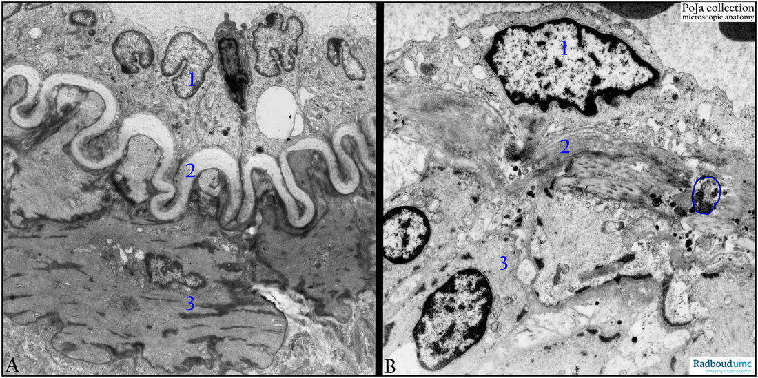
Title: The arteriolar intima in Pseudoxanthoma elasticum (human)
Description:
(A): Electron micrograph of a normal arteriole (example: liver, mouse). (1) Endothelial cells with dense Weibel-Palade granules. The cells are piled up due to contraction of the artery. Between the bulging cytoplasm a squeezed dense-stained lymphocyte. (2) A strong undulated internal elastic lamina. (3) Smooth muscle cells with dense plaques. The surrounding adventitia contains collagen fibrils.
(B): Electron micrograph of an arteriole of a PXE patient. Note that the IEL (2) is fragmented and that ectopic mineralisation with dense Ca-crystal deposits (blue circle) occurs in the collagenous and elastic fibres and components of the ECM. Note the characteristic very dense plaques in the myocytes (3).
Background: Pseudoxanthoma elasticum (PXE) is a disorder characterised by progressive calcification and fragmentation of elastic fibres, and cutaneous lesions. It primarily affects the skin, retina and cardiovascular system. PXE most commonly involves elastic fibres in the reticular dermis of the skin, Bruch membrane in the retina, and smaller/mid-sized arteries. (Ref.: Mohammad J. Hosen, Anouck Lamoen, Anne De Paepe, and Olivier M. Vanakker, “Histopathology of Pseudoxanthoma Elasticum and Related Disorders: Histological Hallmarks and Diagnostic Clues,” Scientifica, vol. 2012, Article ID 598262, 15 pages, 2012. doi:10.6064/2012/598262 http://dx.doi.org/10.6064/2012/598262 ).
Keywords/Mesh: cardiovascular system, vascularisation, blood vessel, arteriole , calcium deposit, smooth muscle, Pseudoxanthoma elastica, electron microscopy, histology, POJA collection