
5.4.2 POJA-L4283+La0089.
Renin in the JG cells adjacent to the glomerulus in kidney V
5.4.2 POJA-L4283+La0089
Title: Renin in the JG cells adjacent to the glomerulus in kidney V
Description:
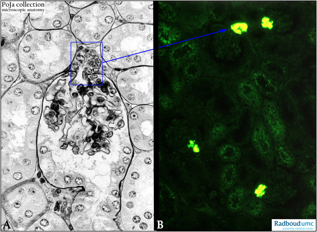
(A): Glomerulus in kidney cortex, silver stain (Movat), mouse. The reticulin fibers and basement membranes are accentuated by this stain.
The blue rectangle focus on the JG cells in the afferent vessel.
(B): Glomeruli in kidney cortex, immunofluorescence staining with anti-renin antibodies, toad. Showing the localization of the renin granules in the JG cells of the wall of the afferent vessel of the glomerulus. Weakly fluorescent dots in the tubules are lysosomes.
(B, by courtesy of A. Lamers PhD, Department Cell biology and Histology, Radboud university medical center, Nijmegen, The Netherlands).
Keywords/Mesh: urinary system, kidney, glomerulus, afferent arteriole, JG cell, renin, histology, POJA collection
Title: Renin in the JG cells adjacent to the glomerulus in kidney V
Description:
(A): Glomerulus in kidney cortex, silver stain (Movat), mouse. The reticulin fibers and basement membranes are accentuated by this stain.
The blue rectangle focus on the JG cells in the afferent vessel.
(B): Glomeruli in kidney cortex, immunofluorescence staining with anti-renin antibodies, toad. Showing the localization of the renin granules in the JG cells of the wall of the afferent vessel of the glomerulus. Weakly fluorescent dots in the tubules are lysosomes.
(B, by courtesy of A. Lamers PhD, Department Cell biology and Histology, Radboud university medical center, Nijmegen, The Netherlands).
Keywords/Mesh: urinary system, kidney, glomerulus, afferent arteriole, JG cell, renin, histology, POJA collection