
5.4.3 POJA-L2418+2432+
4267.
Collecting ducts (XIII) in the human kidney
5.4.3 POJA-L2418+2432+4267
Title: Collecting ducts (XIII) in the human kidney
Description:
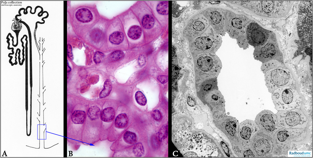
(A): Scheme nephron. The blue rectangle points to a papillary duct (the collecting duct segment, detailed in (B, C).
(B): Stain hematoxylin-eosin, medulla. Inner medullary collecting duct (IMCD). Note also the cross-sectioned capillaries (with red erythrocytes) and intermediate tubules.
(C): Electron micrograph of an OMCD in the cortex. Note that the collecting duct comprises two types of cells, the clear cells (or principal cells) and the dark cells (or intercalated cells). The intercalated are richer in cytoplasmic organelles and possess numerous mitochondria and microvilli (in scanning electron microscopy shown as microridges).
Keywords/Mesh: urinary system, kidney, papillary duct, collecting duct, ductus colligens, dark cell, clear cell, histology, electron microscopy, POJA collection
Title: Collecting ducts (XIII) in the human kidney
Description:
(A): Scheme nephron. The blue rectangle points to a papillary duct (the collecting duct segment, detailed in (B, C).
(B): Stain hematoxylin-eosin, medulla. Inner medullary collecting duct (IMCD). Note also the cross-sectioned capillaries (with red erythrocytes) and intermediate tubules.
(C): Electron micrograph of an OMCD in the cortex. Note that the collecting duct comprises two types of cells, the clear cells (or principal cells) and the dark cells (or intercalated cells). The intercalated are richer in cytoplasmic organelles and possess numerous mitochondria and microvilli (in scanning electron microscopy shown as microridges).
Keywords/Mesh: urinary system, kidney, papillary duct, collecting duct, ductus colligens, dark cell, clear cell, histology, electron microscopy, POJA collection