
11.5 POJA-L3031+3037+
3029+3036+2970+3081.
Oligodendrocytes in cerebrum
11.5 POJA-L3031+3037+3029+3036+2970+3081
Title: Oligodendrocytes in cerebrum
Description:
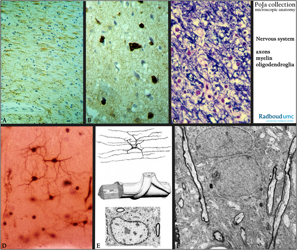
(A): Cortex, human. Immunoperoxidase staining with DAB and antibodies against neurofilament (NF) located in nerve cells and their axons.
(B): Immunoperoxidase staining with DAB and antibody against carboanhydrase, human. Several oligodendroglia cells are stained heavily positive, while small pyramidal cells remain negative.
(C): Stain Kluver-Barrera, human. In the medulla the nuclei of the oligodendrocytes are the dark and smaller ones compared to the other astroglial cells like astrocytes and one microglial cell in diapedesis.
(D): Golgi stain, human. Highlighting the spider-like glia cells (oligodendroglia), schematically detailed in (E).
(E): Electron microscopy scheme of a cell of the oligodendroglia type (human) characterized by having a fine network of “parallel” running delicate processes located between the rows of axon bundles. It forms myelin sheaths around the nerve fibers by wrapping several times around the nerve fibers.
(F): Electron microscopy, rat. Survey neuropil with myelinated (electron-dense rim) and unmyelinated fibers and two small neurons.
The presence of electron-dense melanin is characteristic for nerve cells in the substantia nigra.
Background: Oligodendrocytes ensheath axons to insulate but also to induce sodium channels clustering at the node
of Ranvier, which is important for the salutatory nerve conduction. But oligodendrocytes can also provide trophic support for neurons by production of neurotrophic factors such as glial cell line-derived neurotrophic factor (GDNF), brain-derived neurotrophic factor (BDNF) or insulin-like growth factor-1 (IGF-1).
(A, B, by courtesy of H. ter Laak PhD, Department of Pathology, University Medical Centre Radboud University, Nijmegen, The Netherlands).
Keywords/Mesh: nervous tissue, oligodendrocyte, myelin, carboanhydrase, neurofilament, histology, electron microscopy, POJA collection
Title: Oligodendrocytes in cerebrum
Description:
(A): Cortex, human. Immunoperoxidase staining with DAB and antibodies against neurofilament (NF) located in nerve cells and their axons.
(B): Immunoperoxidase staining with DAB and antibody against carboanhydrase, human. Several oligodendroglia cells are stained heavily positive, while small pyramidal cells remain negative.
(C): Stain Kluver-Barrera, human. In the medulla the nuclei of the oligodendrocytes are the dark and smaller ones compared to the other astroglial cells like astrocytes and one microglial cell in diapedesis.
(D): Golgi stain, human. Highlighting the spider-like glia cells (oligodendroglia), schematically detailed in (E).
(E): Electron microscopy scheme of a cell of the oligodendroglia type (human) characterized by having a fine network of “parallel” running delicate processes located between the rows of axon bundles. It forms myelin sheaths around the nerve fibers by wrapping several times around the nerve fibers.
(F): Electron microscopy, rat. Survey neuropil with myelinated (electron-dense rim) and unmyelinated fibers and two small neurons.
The presence of electron-dense melanin is characteristic for nerve cells in the substantia nigra.
Background: Oligodendrocytes ensheath axons to insulate but also to induce sodium channels clustering at the node
of Ranvier, which is important for the salutatory nerve conduction. But oligodendrocytes can also provide trophic support for neurons by production of neurotrophic factors such as glial cell line-derived neurotrophic factor (GDNF), brain-derived neurotrophic factor (BDNF) or insulin-like growth factor-1 (IGF-1).
(A, B, by courtesy of H. ter Laak PhD, Department of Pathology, University Medical Centre Radboud University, Nijmegen, The Netherlands).
Keywords/Mesh: nervous tissue, oligodendrocyte, myelin, carboanhydrase, neurofilament, histology, electron microscopy, POJA collection