
3.5 POJA-L91AB.
Scheme of tooth development (survey of tooth germ, bell stage)
3.5 POJA-L91AB
Title: Scheme of tooth development (survey of tooth germ, bell stage)
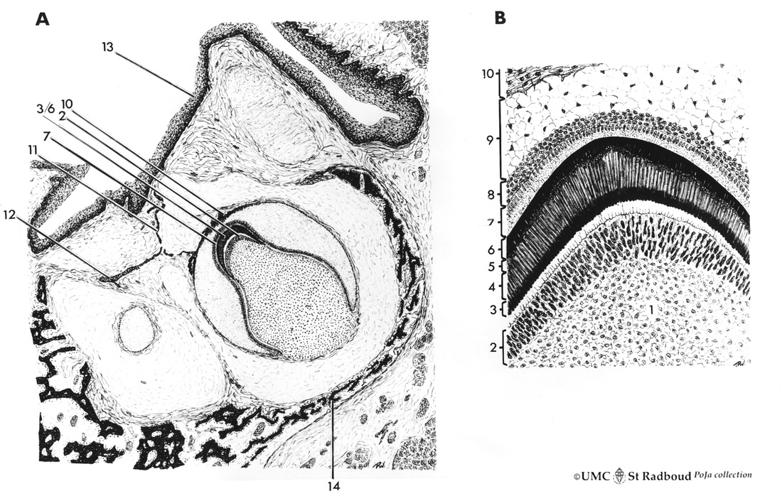
A. Survey of tooth germ. B. Formation of enamel and dentin
Description:
Human embryo.
1. pulp organ 8. stratum intermedium
2. odontoblasts 9. stellate reticulum
3. mantle predentin 10. outer dental epithelium with mesenchyme
4. dentin 11. dental lamina
5. enamel 12. primordium of permanent tooth
6. initial enamel 13. non-keratinized squamous epithelium of oral cavity
7. inner dental epithelium (ameloblasts) 14. alveolar bone (mandible)
Keywords/Mesh: oral cavity, mouth, tooth, tooth development (bell stage), enamel, predentin, dentin, dental lamina, ameloblast, odontoblast, histology, embryology, POJA collection
Title: Scheme of tooth development (survey of tooth germ, bell stage)
A. Survey of tooth germ. B. Formation of enamel and dentin
Description:
Human embryo.
1. pulp organ 8. stratum intermedium
2. odontoblasts 9. stellate reticulum
3. mantle predentin 10. outer dental epithelium with mesenchyme
4. dentin 11. dental lamina
5. enamel 12. primordium of permanent tooth
6. initial enamel 13. non-keratinized squamous epithelium of oral cavity
7. inner dental epithelium (ameloblasts) 14. alveolar bone (mandible)
Keywords/Mesh: oral cavity, mouth, tooth, tooth development (bell stage), enamel, predentin, dentin, dental lamina, ameloblast, odontoblast, histology, embryology, POJA collection

3.5 POJA-L152.
Predentin formation at the cuspal tip in tooth development (bell stage)
3.5 POJA-L152
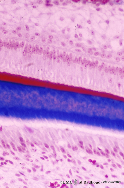
Title: Predentin formation at the cuspal tip in tooth development (bell stage) Description: Stain Azan, human embryo. From top to bottom: - Stellate reticulum consisting of a network of ectoderm-derived cells. - Cell layers of the stratum intermedium. - Columnar (presecretory) ameloblasts with their upper side (nuclear area) in close contact with the stratum intermedium, and at the distal side (secretion area) oriented towards predentin (blue). - Odontoblasts in a epithelioid arrangement with their secretion area close to the predentin (blue). - In close contact with the odontoblasts a network of fibroblast-like cells (so- called pulpal cells) of the dental papilla (future pulp); blood vessels are also present. |

3.5 POJA-L154.
Advanced bell stage with ameloblasts and odontoblasts in tooth development
3.5 POJA-L154
Title: Advanced bell stage with ameloblasts and odontoblasts in tooth development Description: Stain Azan, human embryo. From top to bottom: - Stellate reticulum consisting of a non- vascularized network of ectoderm- derived cells. - Cell layers of the stratum intermedium. - Columnar (presecretory) ameloblasts with their upper side (nuclear area) in close contact with the stratum intermedium, and at the distal side (secretion area) oriented towards predentin (blue). - Predentin with Korff’s fibers. - Tall columnar odontoblasts in a epithelioid arrangement with their secretion area close to the predentin (blue). - Network of mesenchymal derived fibroblast-like cells (so-called pulpal cells) in the dental papilla (future pulp); note blood vessels. |
3.5 POJA-L164.
Ameloblasts and odontoblasts in tooth development (bell stage)
3.5 POJA-L164
Title: Ameloblasts and odontoblasts in tooth development (bell stage) Description: Stain Azan, human embryo. From top to bottom: - Stellate reticulum consisting of a loose network of ectoderm-derived cells. - Cell layers of the stratum intermedium. - Palisade-arranged tall columnar secretory ameloblasts with their nuclear area close to the stratum intermedium. The distal side of the cell is oriented towards initial enamel containing amelogenin (red). - Dentin (blue). Note collagen fibers in the partly decalcified dentin. - Columnar odontoblasts in a epithelioid arrangement with their thin extensions into the predentin (due to preparation procedures the apices of the odontoblasts are detached from the cell bodies). - Fibroblast-like cells (so-called pulpal cells) form the pulp organ, previously dental papilla. |