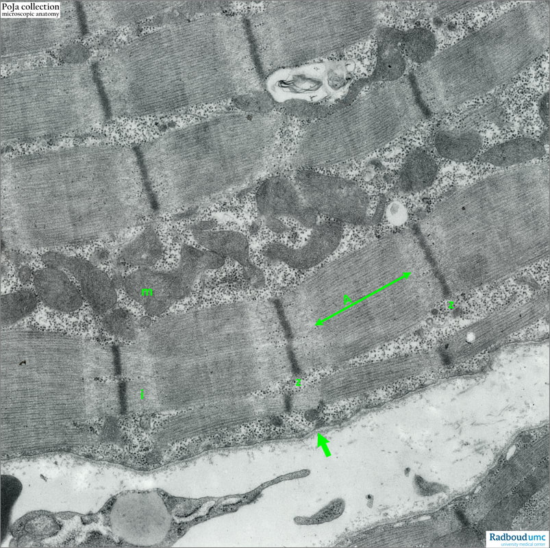
14.1 POJA-L6055 Electron micrograph striated muscle (mouse)
|
14.1 POJA-L6055 Electron micrograph striated muscle (mouse)
Title: Electron micrograph striated muscle (mouse) Description: The sarcomere structure runs from Z- to Z-line. On both sites of the Z-line the less dense stained I-bands exist, while the broad denser stained band is called A-band (anisotropic). The mitochondria (m) are usually interspersed between bundles of myofilaments. The arrow indicates the sarcolemma plus basal lamina. The dark dots between the mitochondria represent the ribosomes and glycogen. Vacuoles with concentric lamellar figures might be present as a kind of autophagic vacuoles or membrane breakdown. Keywords/Mesh: locomotor system, skeletal muscle, striated muscle, myofibre, A-band, I-band, Z-line, mitochondrion, ribosomes, glycogen, electron microscopy, POJA collection |

