4.3.1 POJA-L3808+3809
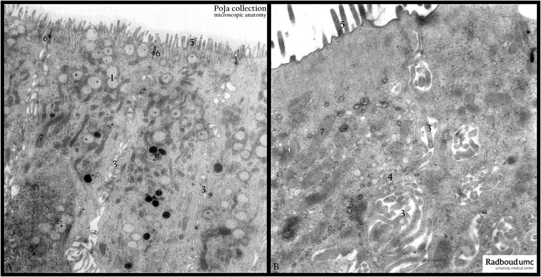
Title: Gallbladder epithelium (monkey, gerbil)
Description: Electron micrographs of gallbladder epithelium of monkey (A) and gerbil (B).
The epithelial cells of the bladder of two different species contain mucus secretion granules (1, 4) used to protect the outside surface of the cells against the concentrated gall liquid.
(2) Electron-dark stained lysosomes.
(3) The intercellular surface of the cells is amplified with microvilli in order to increase water transport capacity of the cells to concentrate the gall liquid in the bladder lumen.
(4) Vesicles.
(5) Microvilli at the lumenal surface.
(6) Note the junctional complexes between cells.
(7) Numerous long slender mitochondria to provide the energy for the water transport and concentration processes.
Keywords/Mesh: gallbladder, water transport, electron microscopy, histology, POJA collection

