
9.3 POJA-L2800+2811+3505
+3503.
Calcitonin cells in thyroid gland (IX)
9.3 POJA-L2800+2811+3505+3503
Title: Calcitonin cells in thyroid gland (IX)
Description:
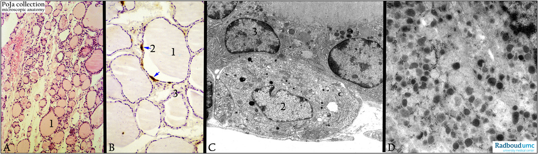
Thyroid gland. (A): Stain hematoxylin-eosin, human. (B): Immunoperoxidase-DAB staining with anti-calcitonin antibodies, human.
Within the follicles (1) of the thyroid gland the inactive hormone product thyroglobulin is temporarily stored.
Most of the lining cells are follicle epithelial cells, but some cells below the lining cells are calcitonin-producing cells demonstrated
in (B, 2 arrows) with the immunoperoxidase reaction.
(C): Electron microscopy, gerbil. (D): Electron microscopy, golden hamster. The parafollicular cells (C, 2 and D) are located just below the normal follicular cells (C, 3), and contain numerous mitochondria and calcitonin-granules which appear here as some small electron-dense and many electron-grey granules (due to partial granule dissolution). The follicular cells (C, 3) are provided with a rich RER profile, basal invaginations and apically larger globules (colloid vacuoles with processed thyroglobulin), small electron-dense lysosomes and lipofuscin. Detail of the cell (D) as in (C, 2) but from a gold hamster.
Keywords/Mesh: thyroid gland, follicle, follicular cell, parafollicular cell, calcitonin cell, C-cell, calcitonin, thyroglobulin, histology, electron microscopy, POJA collection
Title: Calcitonin cells in thyroid gland (IX)
Description:
Thyroid gland. (A): Stain hematoxylin-eosin, human. (B): Immunoperoxidase-DAB staining with anti-calcitonin antibodies, human.
Within the follicles (1) of the thyroid gland the inactive hormone product thyroglobulin is temporarily stored.
Most of the lining cells are follicle epithelial cells, but some cells below the lining cells are calcitonin-producing cells demonstrated
in (B, 2 arrows) with the immunoperoxidase reaction.
(C): Electron microscopy, gerbil. (D): Electron microscopy, golden hamster. The parafollicular cells (C, 2 and D) are located just below the normal follicular cells (C, 3), and contain numerous mitochondria and calcitonin-granules which appear here as some small electron-dense and many electron-grey granules (due to partial granule dissolution). The follicular cells (C, 3) are provided with a rich RER profile, basal invaginations and apically larger globules (colloid vacuoles with processed thyroglobulin), small electron-dense lysosomes and lipofuscin. Detail of the cell (D) as in (C, 2) but from a gold hamster.
Keywords/Mesh: thyroid gland, follicle, follicular cell, parafollicular cell, calcitonin cell, C-cell, calcitonin, thyroglobulin, histology, electron microscopy, POJA collection