
2.1 POJA-L915. Thymus cortex (rat, young adult)
2.1 POJA-L915
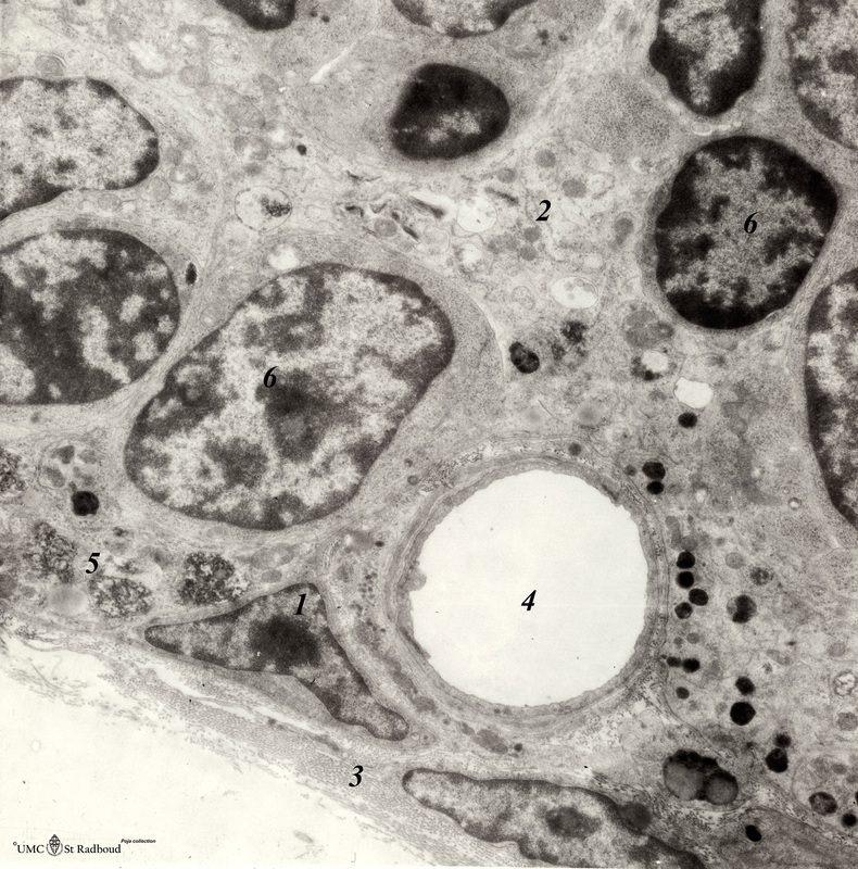
Title: Thymus cortex (rat, young adult)
Description: Electron microscopy.
Type I epithelioreticular cells separate connective tissue compartment from the thymic parenchyma. With occludens junctions and desmosomes as barriers they form wide-mesh networks creating specific microenvironments for developing T cells. The type I cells (1) are located between the connective tissue capsule (3), trabeculae and the thymic parenchyma. The same cells surround the adventitial tissues of cortical blood vessels (4). Type II epithelioreticular cell (2) is located deeper in the cortex and surrounded by thymocytes, the organelles and granules-containing vacuoles are distributed in the squeezed branched projections. (5) part of a macrophage and (6) thymocytes.
Keywords/Mesh: lymphatic tissue, thymus, epithelioreticular cell type I, epithelioreticular cell type II, histology, electron microscopy, POJA collection