10.2 POJA-L2155+2157+2156+2098+2159
Title: Cells of Langerhans in the epidermis
Description:
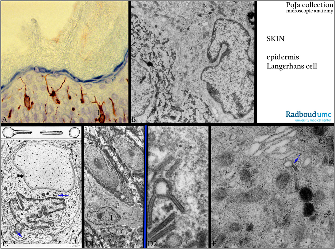
(A): Back skin, immunoperoxidase staining of Langerhans cells (1) with DAB and monoclonal antibodies (LRV202) against
vimentin, human. Langerhans cells are antigen-trapping dendritic cells of the epidermis and involved in cutaneous responses.
The cells of Langerhans are localized in the stratum spinosum with elongated extensions between the keratinocytes.
The vimentin expression, without keratin filaments, confirms their mesenchymal origin.
(B): Back skin, electron micrograph of Langerhans cell (1), human.
(Electron micrograph (B) by courtesy of A. Stadhouders, PhD, Head of the former Department of Electron Microscopy,
Radboud university medical center, Nijmegen, The Netherlands).
(C): Electron microscopy scheme of Langerhans cell, human. (Arrows) Birbeck granule (tennis-racket-shaped structure).
(D1, D2, E): Back skin, electron microscopy, human. Langerhans cell (1) with Birbeck granules (arrow).
Note that in (E) the Birbeck granule is localized close to the Golgi area. The characteristic Birbeck granules (rod-shaped
vermiform granules) surrounded with a membrane, represent endocytic structures that are formed during receptor-
mediated endocytosis. The cells are part of the immunological system, bearing Fc and complement receptors and can present
as well antigens (APC) to naiveT cells.
Keywords/Mesh: skin, epidermis, stratum spinosum, spinous layer, Langerhans cell, dendritic cell, antigen-presenting cell,
Birbeck granule, vimentin,histology, electron microscopy, POJA collection

