11.5 POJA-L3162+3167+3168+3169+3170
Title: Ultrastructure of choroid plexus
Description:
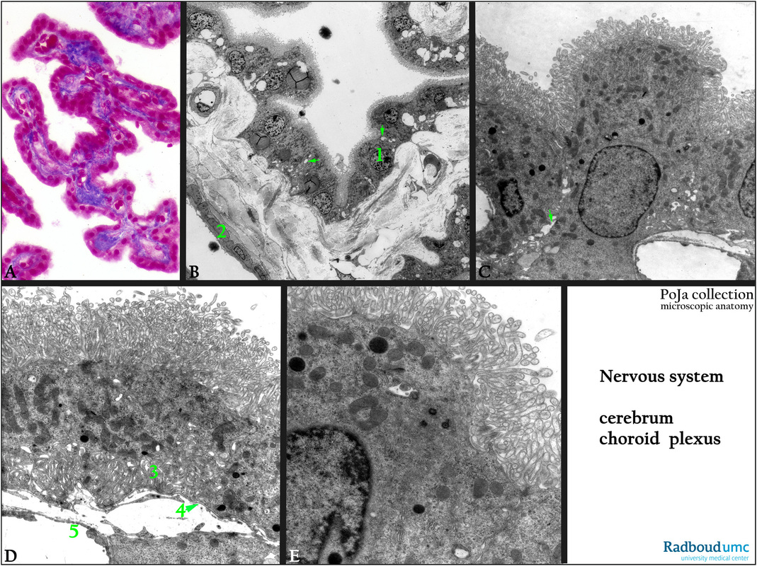
(A): Stain Azan, human. The plexus has a papillary form around connective tissue stalks filled with a capillary network.
It is lined with a single layer of cuboidal epithelial cells being responsible for the production of cerebrospinal fluid (CSF).
(B - E): Electron micrographs of the choroid plexus of a rabbit. The cells (1) are highly polarized. The apical domain contains microvilli,
tight junctions between adjacent cells, and basolateral interdigitating folds. Note also the intercellular spaces between the cells (arrows in B, C) supporting the fluid transport function. (2) Smooth muscle cells of a blood vessel.
The epithelial tight junctions are part of the cerebrospinal fluid (CSF) barrier. The capillaries, however, possess fenestrated endothelium and lack tight junctions. The epithelium of the choroid plexus represents a barrier between the blood and the CSF.
The fenestrated capillary endothelium does not so. Remember that the endotheloid cells of the arachnoid are also linked by tight junctions and thus contribute to the blood-brain barrier as well.
(C): Detail choroidal epithelial cells with numerous small leaf-shaped microvilli, an occasional kinocilium, rabbit.
The epithelial cells are connected by junctional complexes and contain lysosomes.
(D, E): Basal interdigitations (D, 3) in the choroidal cells which are very closely apposed to the capillary with fenestrated endothelium (5), rabbit. Note basal lamina(4) of choroidal cells (zoom in) and apical long microvilli with expanded tips.
Keywords/Mesh: nervous tissue, cerebrum, ventricle, choroid plexus, cerebrospinal fluid, CSF barrier, blood-brain-barrier, histology, electron microscopy, POJA collection

