5.4.2 POJA-La0085+L2343+4429+5024+5029+2487+2488
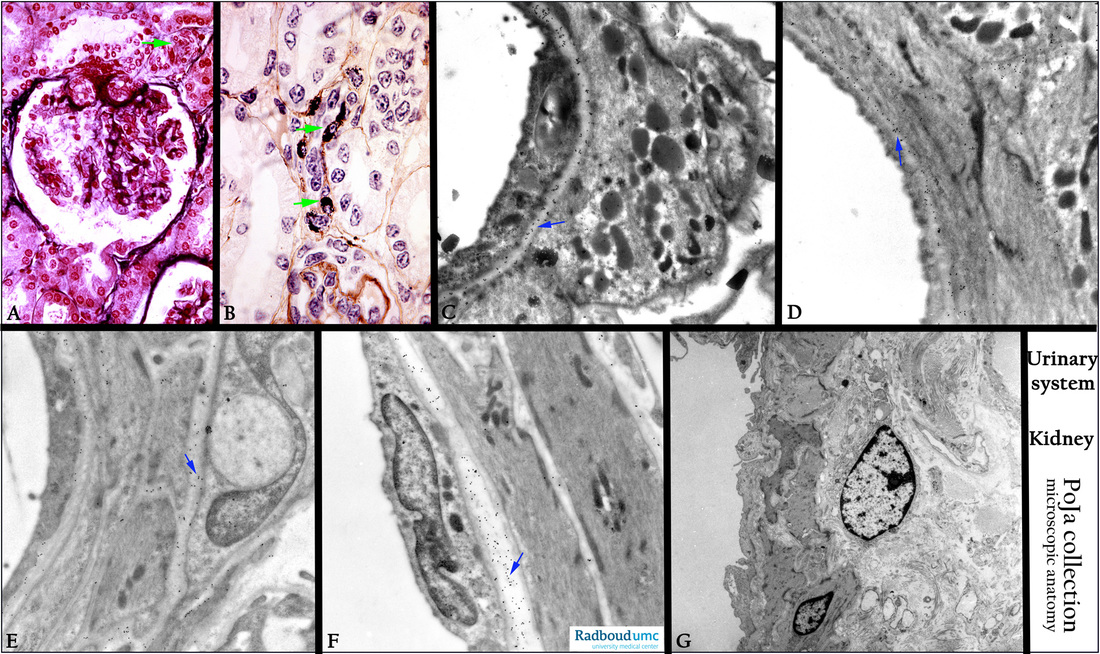
Title: Localization of heparan sulfate (HSPG) in the renal afferent arteriole of the kidney XII
Description:
(A): Survey glomerulus showing darker-stained basement membrane in Bowman’s capsule, and mesangium, stain Azan, human.
(B): Immunoperoxidase staining with DAB and PAP- with IgA antibodies against renin, toad. Compare to (C - F).
Showing the subendothelial localization of granulated cells in the wall (arrows in A, B) of an afferent arteriole of the glomerulus.
Slight background staining due to endogenous Ig in basal lamina and mesangium. (B, by courtesy of A. Lamers PhD, Department Cell biology and Histology, Radboud university medical center, Nijmegen, The Netherlands).
(C-F): Immunogold electron microscopy with antibodies (4C3) against heparan sulfate (arrows), rat. JG granules are negative.
HSPGs is localized in the basal lamina of the vas afferens (a well-organized arteriole) of the glomerulus. Note that the external (basal) lamina of the individual non-granulated smooth muscle cells and the white internal elastic lamina including the basal lamina are HSPG-positive.
(G): Electron microscopy, human. The vas efferens outside the glomerulus is a thin arteriole with less well organized smooth muscle cells surrounded by collagen fibrils and fibroblasts. Note the absence of JG cells.
Keywords/Mesh: urinary system, kidney, glomerulus, vas afferens, heparan sulfate, JG cell, vas efferens, histology, electron microscopy, POJA collection

