
12.2.4.1 POJA-L2962
Inner ear, cochlea I
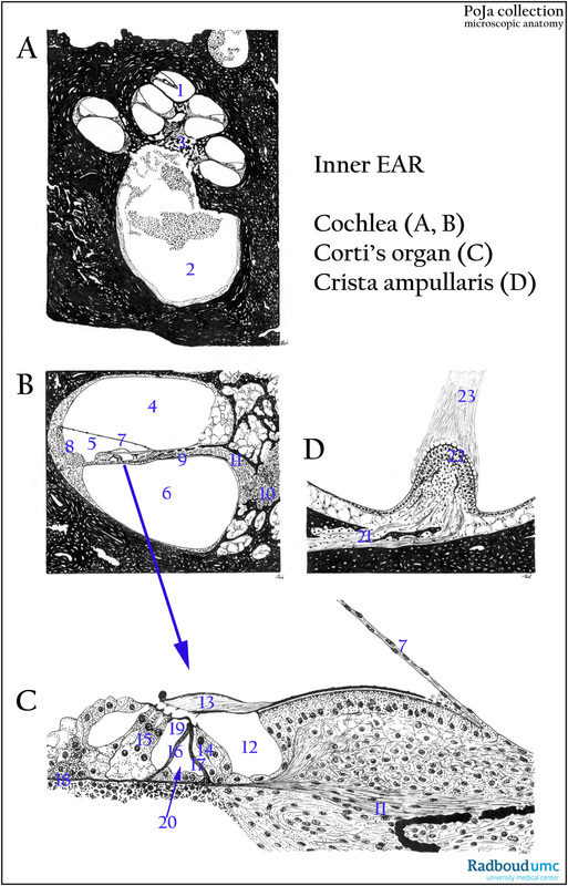
12.2.4.1 POJA-L2962
Title: Inner ear, cochlea I
Description:
Black white drawings of slides (human).
(A): Cochlea.
(1) Helicotrema.
(2) Meatus acusticus internus (or internal auditory meatus) is a channel located in the petrous part of the temporal bone through
which are running: the facial nerve (7th cranial nerve), the vestibulocochlear nerve (or acoustic nerve, auditory nerve,
8th cranial nerve) and the labyrinthine artery (as an internal auditory branch of the basilar artery).
(3) ganglion spirale.
(B): Detail cochlear duct.
(4) Scala vestibuli (perilymphe). (8) Stria vascularis.
(5) Ductus cochlearis (endolymphe). (9) Lamina spiralis ossea.
(6) Scala tympani (perilymphe). (10) Ganglion spirale.
(7) Reissner’s membrane. (11) Fibres of the vestibulocochlear nerve (pars cochlearis).
(C): Organ of Corti.
(7) Reissner’s membrane. (16) Outer pillar cell.
(11) Fibres of the vestibulocochlear nerve (pars cochlearis). (17) Inner pillar cell.
(12) Internal spiral tunnel. (18) Lamina basilaris.
(13) Membrana tectoria. (19) Nuel’s space.
(14) Inner hair cell on inner phalangeal cell. (20) Inner tunnel (tunnel of Corti).
(15) Outer hair cells on outer phalangeal cells.
(D): Ampulla of semicircular canal: crista ampullaris with cupula.
(21) Myelinated nerve fibres of the acoustic nerve.
(22) Crista
(23) Cupula.
Keywords/Mesh: inner ear, cochlea, cochlear duct, organ of Corti, semicircular canal, ampulla, crista ampullaris, histology, POJA collection