16.1.3 POJA-L7086B Osteocyte 1
16.1.3 POJA-L7086B Osteocyte 1
Title: Osteocyte 1
Description:
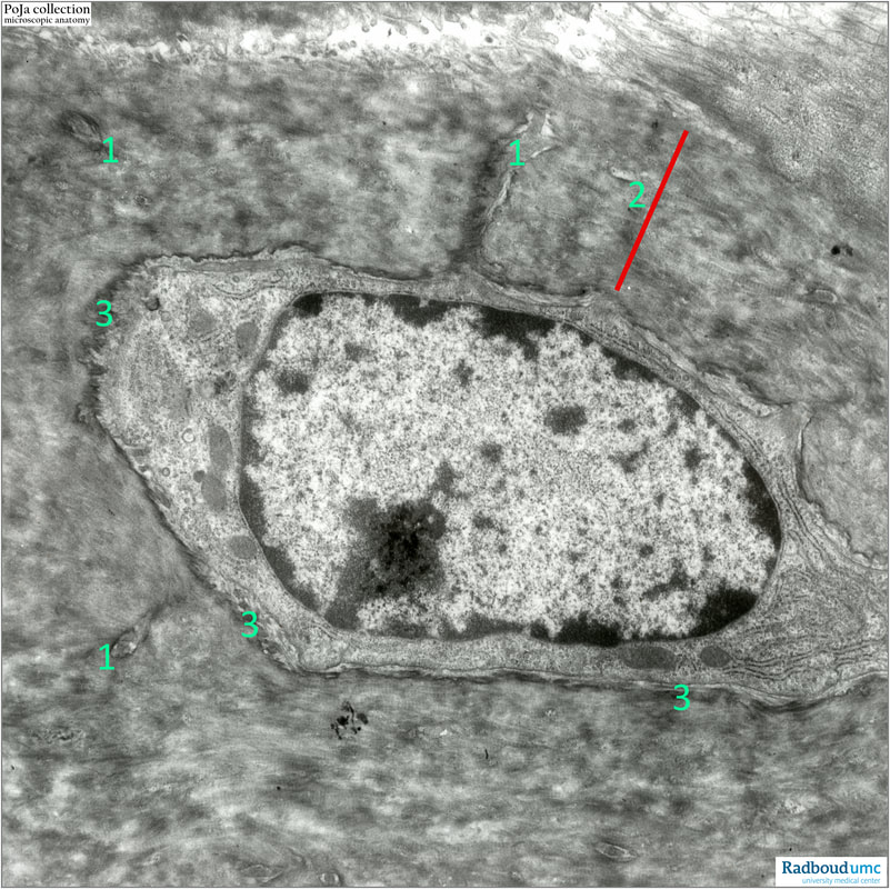
Electron micrograph of a quiescent osteocyte of a rabbit in the process of desmal ossification. Note the nucleus with a conspicuous nucleolus. The cell is located in a lacuna with canaliculi (1). The cell is embedded in a layer of collagen fibrils (2). No hydroxyapatite crystals are present due the decalcifications procedures for the required technical implementation for electron microscopy. The osteocyte is regarded to be in a post-quiescent stage, it has still the distinct osmiophilic lamina (3) close to the cell membrane but the amount of mitochondria and RER profiles appears distinct.
See also: Keywords/Mesh: locomotor system, bone, desmal ossification, osteocyte, lacuna, canaliculi, osmiophilic lamina, collagen fibril, electron microscopy, POJA collection

