13.1 POJA-L4591+4595+4598+4665+4664+4706
Title: Arteries and veins: ultrastructure of surface of endothelial cells
Description:
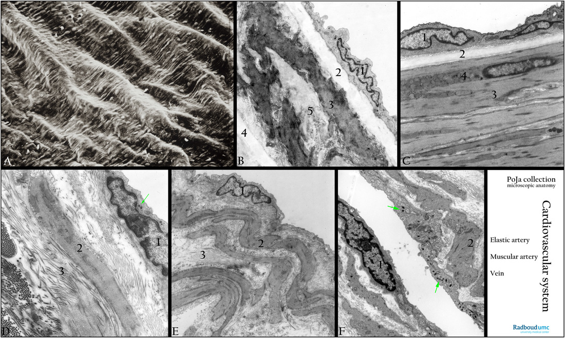
(A): SEM endothelium common carotid artery, gerbil. Inner surface of the blood vessel. The membranous wall of the prepared blood vessel shows imprints of external surrounding structures i.a. bulging thick bundles of collagen. Distinct nuclei of the lining endothelial cells are shown protruding above the surface. Their length axis parallels the axis of blood flow direction.
(B): Mid-sized elastic artery, branch of carotid artery, electron microscopy (EM), rat. (1) Endothelial cell lining the lumen. (2) Internal elastic lamina, all elastic laminae are fenestrated. (3) Branched smooth muscle cells (SMC) with dense plaques. (4) Elastic lamina, or the first elastic lamella which is structurally comparable to the IEL. (5) Mixture of elastic tissue and collagen fibrils is located between the SMC branching processes.
(C): Mid-sized muscular artery, branch of bronchial artery, EM, golden hamster. Similar to (B). Note the accumulation of mitochondria (4) at the nuclear pole in the myocytes.
(D): Electron micrograph of a venule in bronchial region, golden hamster. (1) Endothelial cell with Weibel-Palade granules (arrow). (2) Smooth muscular cell. (3) Collagen and elastic fibers (fibrillogranular material).
(E): Small vein (bronchial region), EM, golden hamster. Intima consisting of endothelial cell (1) and basal lamina. In the media circular layers of SMCs (2) alternate with those of collagen fibrils (3) which are 2-D oriented. Deeper in the wall the amount of collagen fibrils increases. Note that the SMCs are branched.
(F): Electron micrograph of endothelium cell (1) in a small vein, (muscle biopsy, human). Weibel-Palade bodies (arrows). (2) Smooth muscle cells with dense plaques.
Background: The primary function of the lining cells (= endothelium) of a blood vessel is the exchange of substances between blood and tissues. The Weibel-Palade bodies are specific for endothelial cells and appear as elongated organelles (0.1-0.3 micrometers, length 1-5 micrometers formed by the Golgi complex). They represent secretory bodies containing von Willebrand factor (vWF, clotting factor VIII) and proteins. The vWF is stored inside the Weibel-Palade bodies as tubules with a diameter of 12 nm. After release by exocytosis the vWF forms long strings that arrest bleeding by recruiting blood platelets to sites of vascular injury. (Functional architecture of Weibel-Palade bodies. Karine M. Valentijn, J. Evan Sadler, Jack A. Valentijn, Jan Voorberg, Jeroen Eikenboom. Blood May 2011, 117 (19) 5033-5043; DOI: 10.1182/blood-2010-09-267492 http://www.bloodjournal.org/content/117/19/5033?variant=long&sso-checked=true ).
Keywords/Mesh: cardiovascular sytem, vascularisation, blood vessel, artery , vein, Weibel-Palade body, von Willebrand factor, factor VIII, electron microscopy, histology, POJA collection

