
12.1.3 POJA-L2553+4410
Pigmentation of the iris
12.1.3 POJA-L2553+4410
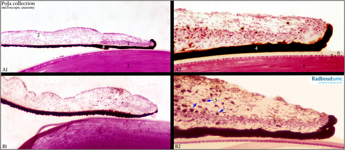
Title: Pigmentation of the iris
Description:
(A, B): Iris and lens, in low and higher magnification, stain Haematoxylin-eosin (variation in staining procedures), human.
(A1) Due to the lack of pigmented melanocytes in the stroma of the iris this eye appears as a blue eye, while (A2) appears darker
and ( B1, B2) manifest themselves as brown eyes due to the numerous pigment-loaded cells in the dense stroma of the iris.
(1) Lens.
(2) Iris.
(3) Musculus sphincter pupillae.
(4) Double epithelial layer with pigment (melanin).
(5) Clusters of melanophores ( melanocytes with pigment). Melanin is yellowish-brown to dark brown in the stromal melanocytes,
and black in iris pigment epithelium. Generally condensation of brownish stromal melanin occurs in the thin anterior border layer.
(6) Capsule of the lens.
(7) Epithelial covering of the lens.
Background: In blue eyes there are less stromal melanocytes and hence less pigment. Light that enters the eye easily traverses
the anterior border layer (light pigmented) and stromal iris. Subsequently it is reflected back out of the eye from the pigmented
epithelium. Due to stromal absorption of the long wave lengths the shorter blue waves will return to the observer eyes.
Usually white babies are born blue-eyed because of its pigmentless stroma. This is in contrast to brown eyes where the stroma is
denser and contains much more clusters of melanocytes with melanin pigments. The process in which non-pigmented stromal
components influence eye color is very complex. Most important elements are selective absorption and reflection by biological
molecules such as haemoglobin (in capillaries), stromal and vessel collagens. The color of the iris is due to variable amounts of
eumelanin (brown/black melanins in brown-eyed persons) and pheomelanin (red/yellow melanins in blue-/green-eyed persons)
produced by melanocytes.
Keyword/Mesh: eye, iris, melanin, melanocyte, blue eye, brown eye, histology, POJA collection