2.2 POJA-L979B
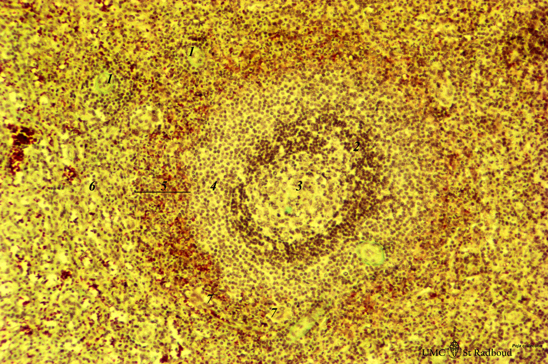
Title: Spleen with secondary lymphatic nodule (human)
Description: Stain: Trichrome (Goldner).
In a more advanced stage the marginal zone (4) develops into a broad zone as shown in this lymph nodule.
Two cross-sections (1) of the central artery surrounded by a small lymphatic sheath (PALS composed of T lymphocytes).
The darker stained mantle zone (2) contains mainly naïve B lymphocytes that encompasses the lighter stained germinal centre (3) filled with reticular cells, B-memory lymphocytes and macrophages. The lighter stained surrounding the mantle zone is the marginal zone (4) (perilymphoid tissue).
The red pulp sinusoids (5) that directly surround the whole lymphoid nodule (as part of the white pulp) contain reddish-stained erythrocytes. The venous sinusoids on a distance appear more empty (6).
At (7) macrophage-sheathed capillaries
Keywords/Mesh: lymphatic tissue, spleen, white pulp, histology, POJA collection

