
11.4 POJA-L4450+3109+
2974+2971+3118+3060.
Purkinje cells in cerebellum 2
11.4 POJA-L4450+3109+2974+2971+3118+3060
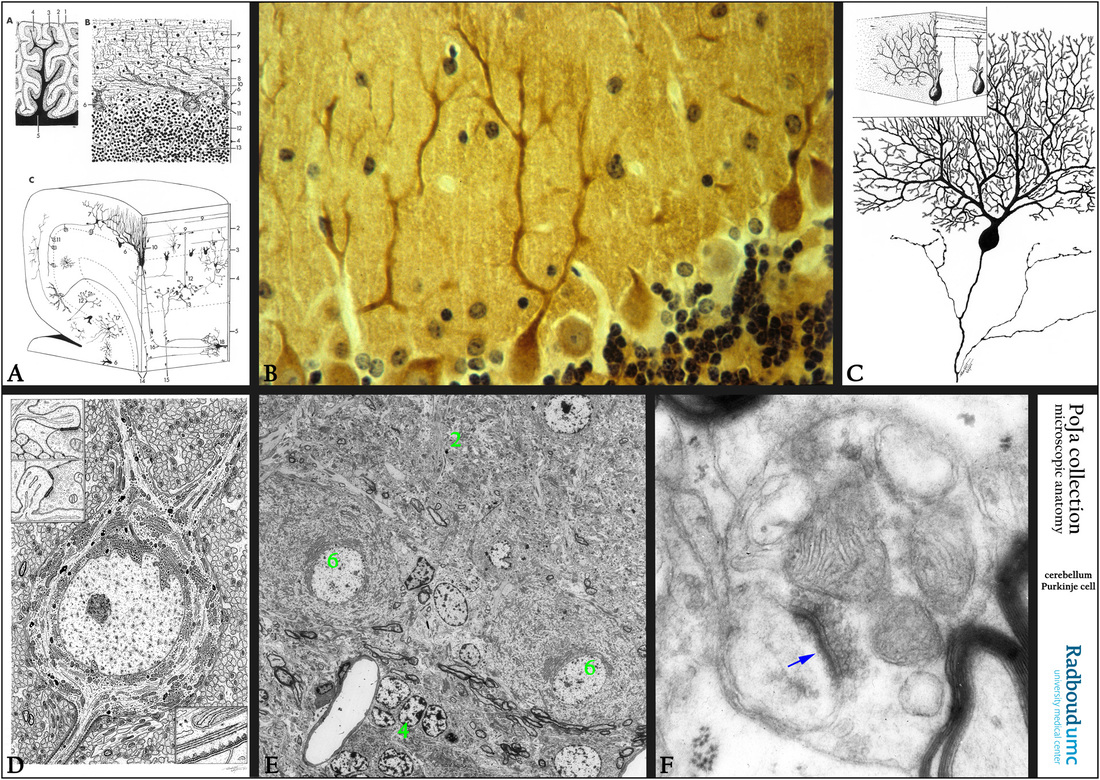
Title: Purkinje cells in cerebellum 2
Description:
A): Scheme cerebellum, human (zoom in).
Referred to Section 11.1 .
(1) Pia mater.
(2) Molecular layer.
(3) Ganglion layer with Purkinje cells.
(4) Granular (or granule) layer.
(5) Medulla rich in myelinated fibers (white matter).
(6) Purkinje cells.
(7) Basket cell (interneuron).
(8) absent in this scheme
(9) Parallel fibers (axons of granule cells).
(10) Dendrites of Purkinje cells in contact with climbing fibers.
(11) Fibers of basket cell around Purkinje cell.
(12) Granule cell (interneuron).
(13) Glomerulus (cerebellar island), contact place between mossy fibers and granule cells.
(14) Climbing fibers (input) ending on the proximal dendrites of Purkinje cells.
(15) Mossy fibers (input) ending in glomerulus.
(16) Axon of Purkinje cell (output from the cerebellar cortex).
(17) Golgi cell (interneuron).
(18) Neuron in deep cerebellar nucleus.
(receive projections from Purkinje cells and send feedback axons to the same cerebellar cortex neurons).
(19) Astrocyte in granular layer. The protoplasmic asterocytes are present in the gray matter and their terminal feet end upon capillaries at one side and thus involved in the blood-brain-barrier (BBB) by formation of perivascular endfeet (membrana gliae limitans perivascularis). At the other side they end up on neurons.
(20) Astrocyte in medulla. These fibrous asterocytes are found in the white matter and just like the protoplasmic asterocytes their processes also
form several perivascular endfeet or subpial endfeet.
(21) Outer stellate cell. The cerebellar stellate neuron is a multipolar interneuron with variable length of their processes and found in the molecular cortex.
(22) Bergmann glial cell. This cell is a specialized semi-radial glia cell that closely interacts with neuronal elements in the molecular layer of the cerebellum. With its long processes it contributes to the formation of the glial limiting membrane.
(23) Lugaro cell. An inhibitory interneuron which appears GABA-positive in the neuritic and somatic profiles. These cells seem to exert a feed-back inhibitory control on Purkinje cells.
(B): Immunoperoxidase staining with DAB and antibodies against tubulin, sagittal section, mouse. Neurotubuli are present in all neuronal structures, the dendritic tree of Purkinje cells shows abundant immunoreactivity.
(C): Scheme of the Purkinje cells with an arbor of ramifying dendrites and one long branched axon, partly sagittal and transversal section (=longitudinal axis of folium of cerebellum), human
(D): Electron microscopy scheme of a large neuron (Purkinje cell), human. Showing a cytoplasm which is rich in Nissl bodies or RER, two dendrites and one neurit, and embedded in neuropil consisting of glia cell, oligodendrocytes and (unmyelinated/myelinated) nerve fibers.
(E): Electron microscopy equivalent of (D), perfusion-fixed, rabbit. (2) Molecular layer. (4) Granule layer. (6) Purkinje cell.
(F): Electron micrograph of a synapse (arrow) in the neuropil, monkey. Note the presynaptic vesicles and a thickened postsynaptic plate.
Keywords/Mesh: nervous tissue, cerebellum, neuropil, Purkinje cell, neuron, Nissl body, axon, dendrite, synapse, synaptic vesicle, neurotubule, histology, electron microscopy, POJA collection