12.2.4.1 POJA-L2624 +La0113+2627+3833
Title: Stria vascularis II in the cochlea of the inner ear
Description:
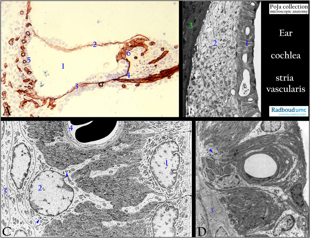
(A): Organ of Corti, immunoperoxidase staining with AEC and antibodies against collagen IV, rat
The cochlear duct (1) is bordered by collagen IV-positive basal laminae, i.e. at the top (Reissner’s membrane, 2), below in the basilar membrane (3), and close to the cochlear nerve fibres (4). The stria vascularis (5) is marked by the positive basal laminae of intraepithelial capillaries. (6) Positive limbus spiralis.
(B): Stain toluidine blue semi-thin plastic section (black and white), detail of stria vascularis, connective tissue of spiral ligament and
part of the bony duct, rat.
Note the intraepithelial vascularisation. The stria vascularis as a specialised secretory, vascular epithelium (1) without a basal lamina,
is separated from the bony cochlear duct (3) by connective tissue (2) of the spiral ligament. The arrangement of the the tight-closed fibroblasts in a dense population with capillaries is characteristic for this specialised form of endosteum of the bony duct.
(C): Electron microscopy scheme of the three layers of the epithelium, human.
(D): Electron micrograph of the epithelium, rat.
(C, D) The stria vascularis consists of (C, 1) marginal cells facing the lumen of the cochlear duct with tight junctions and desmosomen.
These cells possess extensive basal infoldings with numerous mitochondria controlling electrolytes and water transport.
(C, 4) Capillaries with continuous endothelium and basal lamina are present in the middle of the stria closely to the marginal cells.
(C 2, D arrows) Intermediate cells or specialised melanocytes with some melanin granules.
(C, 3; D, 3) Basal cells of this epithelium without lamina basalis closely attached to the underlying spiral ligament fibroblasts.
The stria produces endolymph that resembles intracellular fluid in ionic composition and regulates the K+ homeostasis. But it is also responsible for the maintenance of +90mV endocochlear potential within the cochlear duct. The produced endolymph will be resorbed later on by the endolymphatic sac and transported into the vessels of the dura mater.
With exception of the marginal cells only gap junction connections are present between the intermediate, basal cells and the spiral ligaments cells. Hence the concept of a two-compartment system in the stria for the endolymph and production of generation of endocochlear potential: Na+/ K+-ATPase channels and potassium channels in the intermediate cells and probably also in the basal cells. This implies that the melanocytes are important for the hearing function.
Background: It is assumed that MITF (microphtalmia-associated transcription factor) is a melanocyte-inducible transcription factor.
Mutation of the Mitf gene can cause abnormal skin and iris pigmentation as well as hearing impairment due to loss of melanocytes.
Mitf gene and MITF might be involved in the mitoses in skin- and stria-melanocytes.
Stimuli such as noise increase results in melanogenesis in the intermediate cells of the stria. This is in parallel with the phenomenon of augmentation of the mitosis in skin melanocytes after stimulation e.g. UV radiation, wounding.
Keywords/Mesh: inner ear, cochlea, stria vascularis, marginal cell, melanocyte, capillary, endolymph, K+ homeostasis, histology,
electron microscopy, POJA collection

