
13.1 POJA-L4619+4620 +4621 Arteriovenous anastomoses – adrenal vascular constrictors (human)
13.1 POJA-L4619+4620 +4621
Title: Arteriovenous anastomoses – adrenal vascular constrictors (human)
Description:
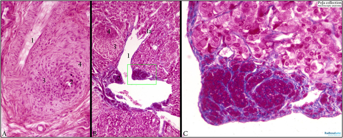
(A): Arterio-venous (AV) anastomosis (subcutis skin), Haematoxylin-eosin. (1) Lumen small artery with common media (cross-sectioned smooth muscle cells or SMC). The same artery is sectioned longitudinally in (1) and partly cross-sectioned in (2). Locally at (3) the arterial wall thickens and transforms into an epithelioid collar of cells involved in the constriction of the vessel. (2) Lumen AV-anastomosis. (3) Epithelioid cells in media. (4) Normal SMCs.
Background (A): Arterio-venous (AV) anastomoses represent direct connections between the arterial and venous system. They are also termed Hoyer–Grosser’s organs or Sucquet-Hoyer corpuscles or glomera cutanea and form thin nodular foci (glomera) 150-200 micrometers, in i.a. pads (stratum reticulare) of fingers, toes, ears, feet. These specialised crossings belong to the arterial constrictors and contain in their media an organised cushing of contractile epithelioid cells. These cells are plump longitudinal arranged modified smooth muscle cells localised between endothelium and media, and there is no internal elastic lamina present. The glomus functions as a shunt- or bypass-regulating mechanism in the flow of blood, indirect control of blood pressure, and temperature and conservation of heat.
(B-C): Specialised vascular constrictors in the adrenal medulla (synonymous to the so-called ‘Drosselvene’), Azan. These specialised vessels may also regulate the blood volume of a capillary network by contraction of the SMCs. They are characterised by longitudinal oriented SMCs in the intima (2). (1) Lumen. (1a) Influx of medullar sinusoids. (2) Intima, cross-sectioned smooth muscle cells. (3) Cortex (fasciculata zone). (4) Medullary cells.
(C): Detail of (B) showing a cushion of smooth muscle cells forming a smooth muscle prominence in the medullary vein.
Background (B): The medullary arterioles of the adrenal gland arise directly from the capsular arteries and form a medullary plexus that drains by small venous vessels into the large medullary vein. The afferent arteriole becomes devoid of the internal elastic membrane, develops a thick epithelioid muscular wall around a small lumen and is connected to short thin-walled veins. Just like the arterial constrictors [in e.g. nasal mucosa (Kiesselbach’s plexus), corpus spongiosum urethrae] SMCs create sphincter-like constructions pushing the blood flow upstream. The innervation occurs by sympathetic, myelinated nerves.
Keywords/Mesh: cardiovascular system, vascularisation, arteriole, vein, vascular constrictor, arteriovenous anastomosis , Hoyer-Grosser’s organ,, glomus cutaneous, histology, POJA collection
Title: Arteriovenous anastomoses – adrenal vascular constrictors (human)
Description:
(A): Arterio-venous (AV) anastomosis (subcutis skin), Haematoxylin-eosin. (1) Lumen small artery with common media (cross-sectioned smooth muscle cells or SMC). The same artery is sectioned longitudinally in (1) and partly cross-sectioned in (2). Locally at (3) the arterial wall thickens and transforms into an epithelioid collar of cells involved in the constriction of the vessel. (2) Lumen AV-anastomosis. (3) Epithelioid cells in media. (4) Normal SMCs.
Background (A): Arterio-venous (AV) anastomoses represent direct connections between the arterial and venous system. They are also termed Hoyer–Grosser’s organs or Sucquet-Hoyer corpuscles or glomera cutanea and form thin nodular foci (glomera) 150-200 micrometers, in i.a. pads (stratum reticulare) of fingers, toes, ears, feet. These specialised crossings belong to the arterial constrictors and contain in their media an organised cushing of contractile epithelioid cells. These cells are plump longitudinal arranged modified smooth muscle cells localised between endothelium and media, and there is no internal elastic lamina present. The glomus functions as a shunt- or bypass-regulating mechanism in the flow of blood, indirect control of blood pressure, and temperature and conservation of heat.
(B-C): Specialised vascular constrictors in the adrenal medulla (synonymous to the so-called ‘Drosselvene’), Azan. These specialised vessels may also regulate the blood volume of a capillary network by contraction of the SMCs. They are characterised by longitudinal oriented SMCs in the intima (2). (1) Lumen. (1a) Influx of medullar sinusoids. (2) Intima, cross-sectioned smooth muscle cells. (3) Cortex (fasciculata zone). (4) Medullary cells.
(C): Detail of (B) showing a cushion of smooth muscle cells forming a smooth muscle prominence in the medullary vein.
Background (B): The medullary arterioles of the adrenal gland arise directly from the capsular arteries and form a medullary plexus that drains by small venous vessels into the large medullary vein. The afferent arteriole becomes devoid of the internal elastic membrane, develops a thick epithelioid muscular wall around a small lumen and is connected to short thin-walled veins. Just like the arterial constrictors [in e.g. nasal mucosa (Kiesselbach’s plexus), corpus spongiosum urethrae] SMCs create sphincter-like constructions pushing the blood flow upstream. The innervation occurs by sympathetic, myelinated nerves.
Keywords/Mesh: cardiovascular system, vascularisation, arteriole, vein, vascular constrictor, arteriovenous anastomosis , Hoyer-Grosser’s organ,, glomus cutaneous, histology, POJA collection