
11.6 POJA-L3874+3875+
3876+3877.
Pathology of degenerative Alzheimer’s disease
11.6 POJA-L3874+3875+3876+3877
Title: Pathology of degenerative Alzheimer’s disease
Description:
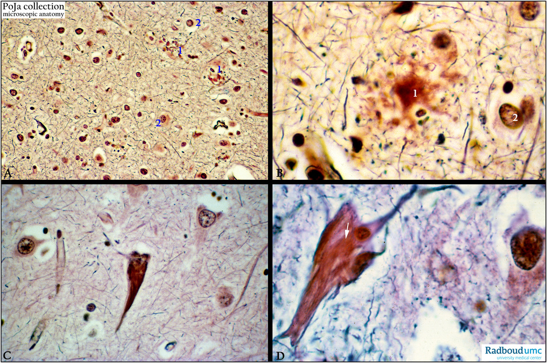
(A-D): Bodian (silver) stain, human. (A, B) Alzheimer plaques (senile plaques) present as neuritic plaque formation with
a rim of tortuous dystrophic neurites surrounding an amyloid core (A1, B1). Reactive astrocytes (A2, B2) and microglia
may appear at the periphery of these plaques. An amyloid precursor protein APP results in fibrillar aggregates of beta-amyloid that is toxic to neurons. Plaques that have the amyloid proteins but lack the neuritic processes are known as diffuse plaques.
(C, D): Neurofibrillary tangle, human. Cells and neurites with the tau protein form so-called tangles. Mostly in cortical neurons
they are shown as basophilic fibrillar structures, these fibrils surround or push away the nucleus and show an elongated ‘flame shape’. The fibrils are composed of paired helical filaments with abnormal hyperphosphorylated tau proteins (arrow).
Other histologic features of Alzheimer disease include amyloid angiopathy with vascular amyloid derived from the same precursor as the amyloid cores of the plaques. Also granulovacuolar degeneration occurs indicated by small intraneuronal cytoplasmic granulated vacuoles. (By courtesy of P. Wesseling MD PhD, Head Neuropathology, Department of Pathology, Radboud university medical center, Nijmegen, The Netherlands).
Background: Additionally to beta-amyloid and tau-proteins a pathological role for a third protein was described in
the Alzheimer’s disease process. The protein TDP-43 correlated with cognitive impairment during life as well as in hippocampal and cortical atrophy. It seems that TDP-43 amplifies memory loss and hippocampal atrophy in Alzheimer’s disease.
Keywords/Mesh: nervous tissue, Alzheimer’s disease, tau, tangle, plaque, pathology, histology, POJA collection
Title: Pathology of degenerative Alzheimer’s disease
Description:
(A-D): Bodian (silver) stain, human. (A, B) Alzheimer plaques (senile plaques) present as neuritic plaque formation with
a rim of tortuous dystrophic neurites surrounding an amyloid core (A1, B1). Reactive astrocytes (A2, B2) and microglia
may appear at the periphery of these plaques. An amyloid precursor protein APP results in fibrillar aggregates of beta-amyloid that is toxic to neurons. Plaques that have the amyloid proteins but lack the neuritic processes are known as diffuse plaques.
(C, D): Neurofibrillary tangle, human. Cells and neurites with the tau protein form so-called tangles. Mostly in cortical neurons
they are shown as basophilic fibrillar structures, these fibrils surround or push away the nucleus and show an elongated ‘flame shape’. The fibrils are composed of paired helical filaments with abnormal hyperphosphorylated tau proteins (arrow).
Other histologic features of Alzheimer disease include amyloid angiopathy with vascular amyloid derived from the same precursor as the amyloid cores of the plaques. Also granulovacuolar degeneration occurs indicated by small intraneuronal cytoplasmic granulated vacuoles. (By courtesy of P. Wesseling MD PhD, Head Neuropathology, Department of Pathology, Radboud university medical center, Nijmegen, The Netherlands).
Background: Additionally to beta-amyloid and tau-proteins a pathological role for a third protein was described in
the Alzheimer’s disease process. The protein TDP-43 correlated with cognitive impairment during life as well as in hippocampal and cortical atrophy. It seems that TDP-43 amplifies memory loss and hippocampal atrophy in Alzheimer’s disease.
Keywords/Mesh: nervous tissue, Alzheimer’s disease, tau, tangle, plaque, pathology, histology, POJA collection