11.2 POJA-L3275+3298+3454
Title: Myelinated peripheral nerve fibers 8
Description:
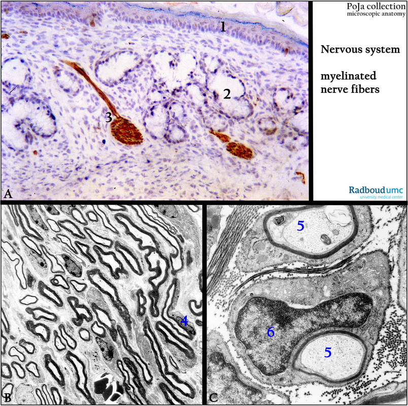
(A): Immunoperoxidase staining with AEC and antibodies against neurofilaments, nerve (A, 3) of the external acousticus meatus,
4d postnatal, rat. (A, 1) Epidermis layers. (2) Glands.
(B): Electron micrograph of a ferrocyanid-contrasted longitudinal section of peripheral nerve fibers, mouse. The myelin sheaths are densely stained. (B, 4) Cell of Schwann.
(C): Electron micrograph of tannin-fixed/stained cross-section through a peripheral nerve, mouse. Two myelinated axons (C, 5) and
a Schwann cell (C, 6) with fuzzy dense-stained basal (external) lamina are shown. The axons contain neurofilaments and neurotubules.
Note longitudinal-/cross-sectioned collagen fibers of the endoneurium.
Keywords/Mesh: nervous tissue, axon, neurofilament, neurotubule, peripheral nerve fiber, myelinated nerve fiber, Schwann cell, endoneurium, histology, electron microscopy, POJA collection

