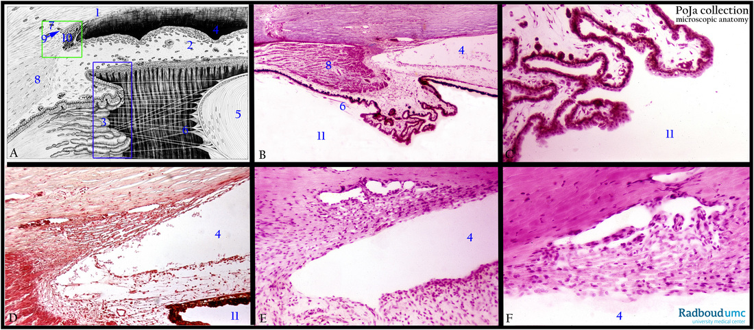
12.1.3 POJA-L4411+2534+4412+4413+2545+2546
Title: Ciliary body and Schlemm canal in the eye
Description:
(A): Ciliary body, electron microscopy scheme, human. (3) Ciliary body. (4) Anterior chamber. (5) Lens, covered with a single
epithelial layer and a capsule of collagen IV and glycosaminoglycans. (6) Suspensory ligament or zonular fibres (or zonules of Zinn,
Zinn’s membrane) arise from the ciliary body (longer fibres and shorter fibres from the pars plana respectively pars plicata).
They form a tent-like structure upon attaching to the lens capsule. They are produced by the non-pigmented epithelial cells of the
ciliary body and contain a.o. collagen type IV as well as fibrillin.
(B, C): Schlemm canal, stain Azan, human. The blue rectangle in (A) represents the ciliary body detailed in (B, C).
Ciliary body and ciliary processes (pars plicata, A, 3) are lined with neuroepithelial cells consisting of a dual layer of
non-pigmented cells and pigmented cells (B, C).
The non-pigmented cells face the posterior chamber (B, C, D, 11) and are continuous with the sensory retina.
The pigmented cells face the stroma of the ciliary processes and are continuous with the retinal pigmented epithelium.
(8) Ciliary muscle.
(D, F): Schlemm canal, stain Haematoxylin-eosin, human. (7) Limbal area with canal of Schlemm (9) and (10) trabecular meshwork of
the corneal-irideal angle. The green rectangle in (A) represents the iridocorneal area with the Schlemm canal system detailed
in (D, E, F). (1) Cornea. Corneal-irideal angle with anterior chamber (A-F, 4) and posterior chamber between lens and ciliary body (11).
(2) Iris with anterior surface without epithelial lining. Loose connective tissue stroma . In the corneoscleral zone a labyrinth of anastomosing channels is called spaces of Fontana (or sinus venosus sclerae of Schlemm canal).
This system drains the chamber water in the anterior chamber and is thus involved in regulating the intra-ocular pressure.
Keywords/Mesh: eye, iris, ciliary body, Schlemm canal, histology, POJA collection

