
12.2.4.1 POJA-La0124+L3403+
3416+2642+2643+2987+3411+2648
Innervation of the organ of Corti in the inner ear
12.2.4.1 POJA-La0124+L3403+3416+2642+2643+2987+3411+2648
Title: Innervation of the organ of Corti in the inner ear
Description:
(A): Survey of the cochlea with the large cochlear nerve bundle (1), stain Azan, guinea pig.
(B): Cochlea, immunoperoxidase staining with AEC and antibodies against collagen IV, rat. Note that all basal laminae
and its collagen IV are well outlined within the nerve bundle (1), around all capillaries and in the limbus area (2).
(C): Spiral ganglion, stain trichrome Goldner, fetus human. Spiral ganglion with maturing neurons most of them still lacking satellite cells.
(D): Spiral ganglion, stain Azan, guinea pig. Ganglion with characteristic bipolar neurons and its nerve fibres between the blue-stained
osseus spiralis lamina.
(E): Spiral ganglion, immunoperoxidase staining with AEC and antibodies against neurofilament, 4 days postnatal rat.
Spiral ganglion containing young maturing neurons with varying positive staining.
(F): Spiral ganglion, immunoperoxidase staining with AEC and antibodies against laminin, 1 day postnatal rat.
Cross-sectioned myelinated axons, capillaries are positively stained.
(G): Spiral ganglion, immunoperoxidase staining with AEC and antibodies against collagen IV, 10 days postnatal rat.
A whole spiral ganglion shows collagen IV of basal laminae around the nerve fibres and blood vessels.
Ganglion cells and cartilage cells of the future osseus spiral lamina are negative.
(H): Cochlear nerve, immunoperoxidase staining with AEC and antibodies against neurofilament, 1 day postnatal rat.
The delicate individual axons of the cochlear nerve containing neurofilament proteins are specifically well-stained.
Keywords/Mesh: inner ear, fetus, cochlea, organ of Corti, spiral ganglion, collagen IV, laminin, neurofilament, histology, POJA collection
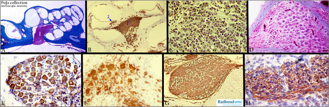