
7.2 POJA-L1221+1228. Fallopian tube, ampulla (human, adult)
7.2 POJA-L1221+1228
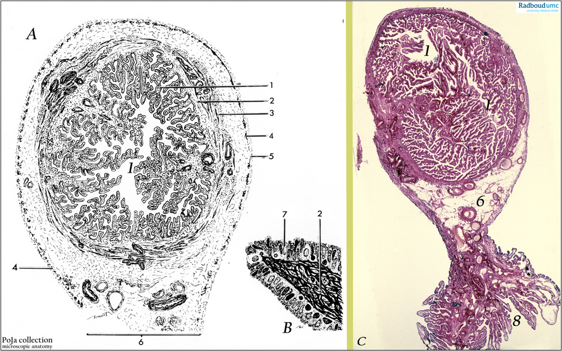
Title: Fallopian tube, ampulla (human, adult)
Description: (A-B) Scheme of ampulla of fallopian tube ; (C) Stain: Hematoxylin-eosin.
(A-B): Scheme:
1. mucosal folds (plicae) (A);
2. proper lamina (A, B);
3. muscular layer (A);
4. sub peritoneal muscles (A);
5. serosa (A);
6. mesosalpinx (A,C);
7. columnar ciliated and secretory cell lining (B);
8. part of infundibulum with fimbriae(C);
(C): Survey of ampulla. Lumen with tortuous major plicae and large numbers of minor plicae.
Background: The uterine tube (fallopian tube or oviduct) is about 10-12 cm long and consists of a muscular tube which lumen is lined with a mucosa. The one end shows finger-like processes that can engulf the ovary. The other end of the tube connects with the uterine lumen. Four parts are discerned:
(1) The infundibulum containing fimbriae or folds with variable processes close upon the ovary.
(2) The ampulla has a length of ca. 7-8 cm and a maximum width of 1 cm. It is the site of fertilization. The ampulla is characterized by tortuously folded mucosal lining with about 4-5 major longitudinal ridges (plicae) on which a large number of secondary plicae are present. (3) The isthmus is ca. 4 cm in length, with a narrow lumen of 0.1 – 0.5 mm. The isthmus is firm and most muscular and possesses ca. 3-5 major plicae with fewer minor plicae.
(4) The short intramural (or intrauterine) part (ca. 0.6 mm in length) is localized within the wall of the uterus ending in the uterine cavity close to upper end of the uterine cornu.
The oviduct is surrounded by an external serosa. The external serosa is actually a peritoneum with subjacent connective tissue continuous with the broad ligament. The strongly folded mucosa provides the lumen a labyrinth-like lining. The mucosa itself consists of a layer of columnar ciliated and secretory cells resting on a proper lamina that is richly supplied by blood vessels and lymphatic capillaries. The plicae produce a giant enlargement of the epithelial cell surface. During the pre-ovulatory period or estrogenic phase the number, the height and the ciliary activity of the ciliated cells increase. In the progesteron phase a decrease of all phenomena occurs including lost of cilia (or so- called deciliation). Though there are less secretory cells than ciliated ones the estrogen effect stimulates secretion activity followed by increased discharge during the luteal phase. The secretion product is thick watery serum-like mucus, rich in potassium, chloride and immunoglobulins. Young secretory cells are also named peg or intercalary cells. Few basally located small cells function as stem cells for both ciliated and secretory cells. Interestingly, a mixed layer of smooth muscle fibers, venous and lymphatic capillaries is localized between mucosa and muscularis. At the time of ovulation this zone gets filled with lymph and blood and subsequently functions as a kind of erectile tissue also known in e.g. vagina, labia minora. In this way the infundibula draw up and the fimbriae will approach closely to the ovaria by the time of ovulation. The released ovum is thus prevented from falling into the peritoneal cavity and will be captured easily by the fimbriae nearly engulfing the ovary. The muscularis consists of circular internal and longitudinal smooth muscle layers which are, however, arranged as tight spirally (circular) and loose spirally (longitudinal) fibers. Recently a new cell-type has been described the so-called interstitial Cajal-like cells. These cells are involved in cell signaling, act as steroid sensors and excitatory regulators of smooth muscle motility. The muscle fibers propagate rhythmic contractions toward the uterus thereby enhancing both the contact between ovum and sperm and the course of the fertilized ovum. In the ampulla and infundibulum the circular and longitudinal muscle fibers are intermingled. The isthmus part, however, possesses thick well defined circular muscles. The intramural part shows no sphincters predominantly internal longitudinal muscle fibers. No muscle sphincter is present. However a zone of vascular tissue loops around the os or aperture of the uterine cavity. Likely this construction contributes in sealing off the oviduct-uterus connection and thus protects from infection..
Keywords/Mesh: female reproductive organs, oviduct, fallopian tube, histology, POJA collection, epithelium, cilia, Cajal-like cells, fimbriae, histology, POJA collection