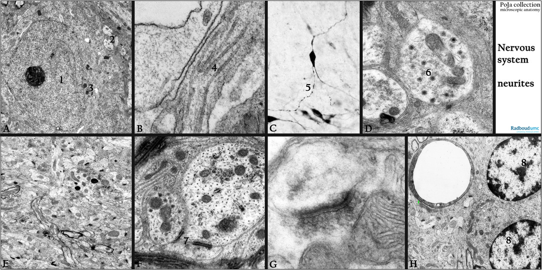
11.5 POJA-L3079+3367+3074+3302+3086+3300+3061+3065
Title: Axonal projections in cerebrum/cerebellum
Description:
(A): Electron micrograph of a neuron (1), axons (2) with neurotubuli. Neurons in the substantia nigra area contain neuromelanin granules (3), rat.
(B): Electron micrograph of an axon with neurotubules (4), fetus, human
(C): Vibratome 60 µm section immunoperoxidase staining with DAB and antibodies against corticotroph releasing hormone (CRH), rat.
The accumulated secretion droplets in the axon in a hypothalamus area (paraventricular nucleus) appear like a string of beads (5).
(Stained section kindly supplied by J. Dederen BSc, Department of Anatomy, University Medical Centre Radboud University, Nijmegen, The Netherlands).
(D): Electron micrograph of neurosecretion granules (6) (e.g. CRH) in an axon, rabbit.
(E): Electron micrograph of neuropil and synapses in corpus striatum, rat.
(F): Electron microscopy, rabbit. Axon-axonal synapse with symmetrical electron-dense zones of the same size in pre- as well as in postsynaptic areas (type 2) (7).
(G): Electron microscopy, monkey. An axon-dendritic synapse with presynaptic transmitter vesicles in cerebellum. An asymmetrical synapse (type 1) with a prominent accumulation of electron-dense material on the postsynaptic side.
(H): Electron microscopy, perfusion-fixed, rat. Neuropil in the corpus striatum. Note the capillary surrounded with a pericyte (arrow)
and adjacent a very small rim of glial cytoplasm. (8) Two small neurons.
Keywords/Mesh: nervous tissue, cerebrum, cerebellum, substantia nigra, hypothalamus, neuropil, neuron, axon, dendrite, neurotubule, synapse, synaptic vesicle, neurotransmitter, corticotroph releasing hormone, neurosecretion, histology, electron microscopy, POJA collection

