11.3 POJA-L3141+3135+3050+3067
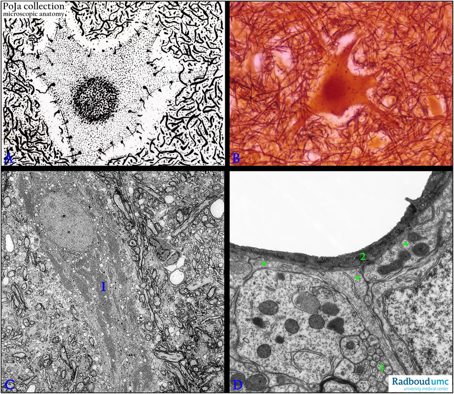
Title: Motoneurons in anterior column of the spinal cord
Description:
(A): Scheme motoneuron, dog. The large multipolar neuron cell body receives numerous neurites, or dendrites
on its cell surface as “boutons terminaux” (terminal buttons).
(B): Silver-stained large motoneuron in anterior column (ventral column), equivalent to (A), dog.
(C): Electron microscopy, rat. Survey of a motoneuron embedded in a network of glia cells and numerous
electron-dense, myelinated axons. Note the Nissl bodies (1) in the perikaryon of the multipolar cell.
(D): Electron microscopy, rat. Blood-brain barrier (BBB) in the posterior column (substantia gelatinosa).
(2) Capillary lined with endothelium (BBB), (**) astrocytic endfeet apposed to the basal lamina of the capillary.
(By courtesy of P. Buma PhD, Department of Orthopedics, University Medical Centre of the Radboud University, Nijmegen,
The Netherlands).
Background: Referred to Section 11.1 The apex of the posterior horn is capped by a V-shaped mass of translucent, gelatinous neuroglia, termed substantia gelatinosa of Roland. The gelatinous appearance is due to a very high concentration of unmyelinated fibers as shown in (D, 3).
Keywords/Mesh: nervous tissue, spinal cord, motoneuron, Nissl body, axon, terminal button, blood-brain barrier, histology, electron microscopy, POJA collection

