
13.1 POJA-L4639+4641+4583+4640+4638+4634a+4637+La0303
Arterioles and small veins
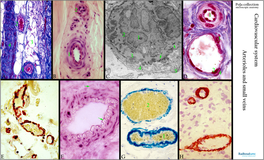
13.1 POJA-L4639+4641+4583+4640+4638+4634a+4637+La0303
Title: Arterioles and small veins
Description:
(A): Azan staining, human (1) Arteriole with 1-2 layers of smooth muscles in the wall. (2) Small vein, thin walled and wide lumen. (3) Adipocytes. (4) Bundle of myelinated nerves. There is no or less developed internal elastic lamina (IEL), almost no media with smooth muscle cells (SMC) in the vein.
(B): Haematoxylin-azophloxine staining (human) of a small-sized artery (1) showing an elastica interna (red) and smooth muscle cells in their wall. Note difference with the contracted arteriole (2) missing a visible IEL.
(C): Electron micrograph arteriole in bronchiolar region, golden hamster. The contracted lumen is lined by endothelial cells (1), which rest on a basal lamina (2), adjacent to the IEL which is discrete and fenestrated. (3) Smooth muscle cells with dense attachment sites (arrows). (4) Nerve cells contain slender myelin-free processes (5) with vesicles. Basal laminae of axon and SMCs might be fused. This image is also demonstrated in peripheral blood vessels: ultrastructure of intima-media 13.1 POJA-L4643+4583+4595+4579+4707+4667.
(D): Azan staining of a small arteriole (1) and a venule (2), skin, human. The media of the arteriole with 1-4 SMCs is followed by a collagenous adventitia fixed to its surroundings. The muscular venule is lined by endothelial cells (2), a single SMC (3), a single pericyte (4) and thin collagenous adventitia.
(E): Immunoperoxidase with AEC staining for α-SM-actin in the wall of veins, submucosa colon, human.
(F): G6PDH (glucose-6-phosphate dehydrogenase) staining in the smooth muscle cells in the wall of muscular artery. Note both the membrane elastica interna and externa (arrows), kidney, mouse.
(G): Immunochemical staining with anti-α-Sm actin-b-galactosidase conjugated antibodies counterstained with haematoxylin, human. Staining for actin in a small artery (1) and a vein (2), submucosa colon, human.
(H): Immunoperoxidase staining with AEC for collagen IV in vascular basement membrane of a vein and 2 small capillaries in the cerebral tissue of rat, 1 day postnat.
Nuclei of neuroglia cells of the neuropil (nervous tissue without neuronal nuclei) are also counterstained with haematoxylin. The two small capillaries possess 1-2 pericytes. Note that all vascular structures are completely sealed off by their positive basement membranes from the neuropil (Blood-Brain Barrier).
Background: Arterioles are the smallest arteries (diameter of 20-120 micrometers) with minimal 1-2 layers of smooth muscle cells (SMC) in the wall. The terminal arterioles (also termed pre-capillaries) are followed by a network of arterial capillaries that drains via post-capillary venules and pericytic c.q. muscular venules into collecting venules. The latter precede small veins (diameter 1-2 mm) followed by mid-sized veins and large veins. Veins have a larger volume and a relatively thinner wall. Although intimal, medial and adventitial layers are present in veins, they are less clearly demarcated than in arteries. Arterioles, capillaries and post-capillary venules together comprise the microcirculation where exchange of products occurs. In the adventitia sympathetic and parasympathetic nerve plexus (nervi vasorum) are located with myelin-free processes.
Keywords/Mesh: cardiovascular system, vascularisation, blood vessel, arteriole , muscular artery, vein, small vein, basal lamina, collagen IV, actin, pericyte, histology, POJA collection