
5.7 POJA-L5015+5016+
5019+5014+5018.
Detrusor muscle in the human urinary bladder 3
5.7 POJA-L5015+5016+5019+5014+5018
Title: Detrusor muscle in the human urinary bladder 3
Description:
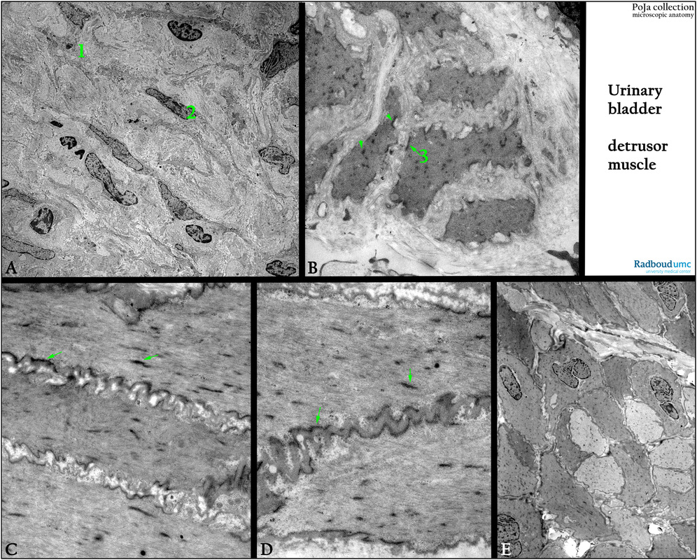
Electron microscopy. (A): Lamina propria of the urinary bladder with fibroblasts and interstitial cells.
(B, C, D, E): Smooth muscle structure of the detrusor muscle of the urinary bladder illustrating the dense plaques (3, arrows, arrowheads) within the muscle cells. (A, 1) The myofibroblast in the lamina propria has a stellate morphology with multiple branches. The cell is accompanied by fibroblasts (A, 2). The detrusor has a compact fascicle (E) with cylindrical to polygonal profile of closely packed smooth muscle cells with numerous dense plaques. Contiguous cells are adjoined by complementary electron dense areas (arrows) at the membrane (C, D).
The undulating (D) cellular membranes indicate incomplete cell relaxation.
Keywords/Mesh: urinary system, urinary bladder, vesica urinaria, detrusor muscle, smooth muscle, dense plaque, histology, electron microscopy, POJA collection
Title: Detrusor muscle in the human urinary bladder 3
Description:
Electron microscopy. (A): Lamina propria of the urinary bladder with fibroblasts and interstitial cells.
(B, C, D, E): Smooth muscle structure of the detrusor muscle of the urinary bladder illustrating the dense plaques (3, arrows, arrowheads) within the muscle cells. (A, 1) The myofibroblast in the lamina propria has a stellate morphology with multiple branches. The cell is accompanied by fibroblasts (A, 2). The detrusor has a compact fascicle (E) with cylindrical to polygonal profile of closely packed smooth muscle cells with numerous dense plaques. Contiguous cells are adjoined by complementary electron dense areas (arrows) at the membrane (C, D).
The undulating (D) cellular membranes indicate incomplete cell relaxation.
Keywords/Mesh: urinary system, urinary bladder, vesica urinaria, detrusor muscle, smooth muscle, dense plaque, histology, electron microscopy, POJA collection