10.4 POJA-L2085+2086+2201+2269+2186+2205+2207
Title: Eccrine sweat glands and myoepithelial cells
Description:
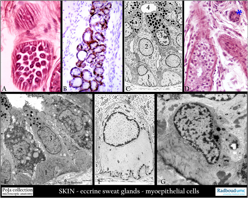
(A): Axillar skin, stain hematoxylin-azophloxine, human. Eccrine sweat glands with tangential cut myoepithelial cells.
(B): Back skin, immunoperoxidase staining with DAB and antibodies (RCK 107) against cytokeratin 14, human.
The cells also contain cytokeratin 17.
(C): Electron microscopy scheme of the duct of the eccrine sweat gland, human.
(1) Myoepithelial cell with dense plaques.
(2) Clear cells rich in glycogen.
(3) Secretory, mucoid epithelial cells with dark secretion granules.
(4) Lumen of the gland.
(D): Axillar skin, stain hematoxylin-azophloxine, human. Eccrine sweat gland, secretory part with double layered epithelium
with clear and dark cells.
(*) Duct of the gland.
(E): Back skin, electron micrograph of eccrine sweat gland, human. Two types of cells are discerned: a dark cell type
with many RER profiles and secretory granules, and a light cell type without RER and glycogen, but instead being well
provided with mitochondria arranged in rows between deep intercellular invaginations (related to ion transport).
(1) Myoepithelial cell.
(2) Clear cell rich in glycogen. This cell secretes water and NaCl.
(3) Dark cells with secretion granules containing glycoprotein.
(F): Electron micrograph scheme of a myoepithelial cell with contractile filaments such as actin, human.
Note the dense plaques.
(G): Back skin, electron micrograph of a myoepithelial cell, human. The secretory portions contain myoepithelial cells
in order to squeeze out the secretory product.
Keywords/Mesh: skin, eccrine gland, myoepithelial cell, cytokeratin, histology, electron microscopy, POJA collection

