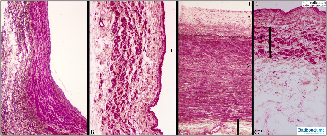
13.1 POJA-L4666+4566+4567
Title: Large veins (human)
Description:
(A): Femoral vein, resorcin-fuchsin and light green. Due to the hydrostatic pressure (below the heart level) adaptations are present in especially veins of the limbs. Longitudinal arranged smooth muscle cells (SMC) appear in the intima and adventitia in different areas of the wall of the vein, the media appears thin and there is a gradual transition into the adventitia. In one section two different thickness of the wall are shown.
The intima (1) with longitudinal arranged SMC. Media (2) with circular bundles of SMC, separated by irregular layers of connective tissue. In the thickest part of the wall the media-adventitia (3) is characterized by partly circular to longitudinal arranged SMC.
(B): Inferior cava vein, resorcin-fuchsin and modified light green. Lumen of inferior cava (1), intima (2) and media (3) ill-delimited from a thick adventitia (4). The intima lined by a thin endothelium might contain fine elastic fibres and an internal elastic lamina. The thin media contains irregular circular orientated smooth muscle cells with loose connective tissue and thin elastic fibres. The transition from media to adventitia is gradual and the border ill-defined. Longitudinal oriented muscular tissue dominates in bundles encircled by collagenous tissue, intermingled with elastic fibres. Surrounding connective tissue (5) with vasa vasorum (6).
(C1-2): Comparison of Aorta (C1) and inferior cava vein (C2) resorcin-fuchsin and light green stain. Lumen of vessel (1). Intima (2). Media (3). Vertical bar indicates adventitia (4). Note the difference between the largest conducting artery (aorta) and the largest vein (inferior cava) in the human body. The media in the artery (C1) is very thick compared to the media of the vein (C2). However, the adventitia of the vein (C2) contains considerable SMC bundles in contrast to the adventitia of the aorta (C1).
Background: Generally large veins possess a relatively thin wall with a well-developed adventitia. The intima contains some subendothelial collagen, elastic fibres and close to the media reinforcement by longitudinal oriented smooth muscle cells (SMC). The larger the veins the lesser circular SMCs in the media and the more longitudinal oriented SMCs in the adventitia. There is almost no sharp delineation between media /adventitia. Vasa vasorum is present in the vessel walls thicker than 70 µm. In varying the length tension of the wall the vessel meets to the changed pressures between lumen and environment of the vein. Even during underpressure the lumen of the vein remains open.
Keywords/Mesh: cardiovascular system, vascularisation, vein, inferior cava vein, femoral vein, smooth muscle cell, hydrostatic pressure, histology, POJA collection

